Muscles on Back Of Neck
Table of Contents
What muscles are in the back of the neck?
The muscles on the Back of the neck are muscles that cover the neck area. The neck muscles are part of a complex musculoskeletal system that connects the base of your skull to your torso. The muscles of the neck are mainly responsible for the movement of the head in all directions.
They consist of 3 main groups of muscles: anterior, lateral, and posterior muscle groups, based on their position in the neck. The muscles of the neck are further divided into more specific groups based on a number of determinants, including depth, precise location, and function.
The position of a muscle or group of muscles in the neck generally relates to the function of the muscles in the neck. For example, the muscles in the posterior compartment of the neck are responsible for the extension of the neck. The muscles of the neck are closely related to a number of important structures that pass between the thorax and the head, which include major blood vessels, nerves, and elements of the respiratory and gastrointestinal systems.
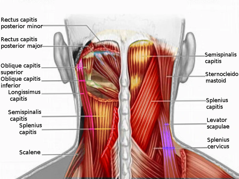
Muscles on the back of the neck include:
- Superficial layer: Trapezius, splenius capitis, splenius cervicis
- Deep layer: Cervical transversospinales muscles
- Semispinalis capitis,
- Semispinalis cervicis
- Deepest layer: Suboccipital muscles:
- Rectus capitis posterior major,
- Rectus capitis posterior minor,
- Obliquus capitis superior,
- Obliquus capitis inferior
The superficial layer of muscles on the back of the neck
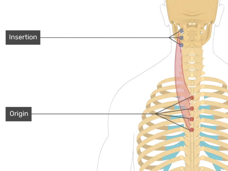
The Splenius Capitis muscle
Origin:
The splenius Capitis originates from the spinous processes of vertebrae C7 to T3 and the lower half of the nuchal ligament.
Insertion :
The splenius Capitis was inserted over the mastoid process and the lateral third of the superior nuchal line of the occipital bone.
Blood supply :
blood supplies The splenius Capitis are the vertebral, occipital, superior intercostal, deep cervical, and transverse cervical arteries.
Nerve supply:
The nerve supply of the splenius Capitis is the posterior rami of the middle cervical spinal nerves and the lower cervical spinal nerves
Action :
The splenius Capitis does lateral flexion of the neck and rotation of the neck to the same side.
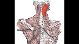
The Splenius Cervices
Origine :
The Splenius Cervicis originate from the spinous processes of vertebrae T3 to T6
Insertion :
The Splenius Cervicis are inserted over the transverse processes of vertebrae C1 to C3 or C4.
Blood supply :
The blood supply of the splenius Cervicis is the vertebral, occipital, superior intercostal, deep cervical, and transverse cervical arteries.
Nerve supply :
The splenius Cervicis muscle’s nerve supply is the posterior rami of the middle cervical spinal nerves and lower cervical spinal nerves
Action :
The Splenius Cervicis do lateral flexion of the neck and rotation of the neck to the same side.
Trapezius muscle
Origin :
The Trapezius muscle originates from the medial third of the superior nuchal line; external occipital protuberance, nuchal ligament, and spinous processes of C7 to T12 vertebrae.
Insertion :
The trapezius muscle inserts on the lateral third of the clavicle, acromion, and spine of the scapula.
Nerve Supply:
The Trapezius muscle’s nerve supply is the spinal root of the accessory nerve (CN XI) (motor)
C3 and C4 Cervical nerves (pain and proprioception)
Blood Supply:
The Trapezius muscle’s arterial supply is the Transverse cervical artery (cervicodorsal trunk)
Action :
The function of the trapezius muscles is to stabilize and move the scapula. The upper fibers of the trapezius muscles elevate and upwardly rotate the scapula and extend the neck. The middle fibers of the trapezius muscles adduct (retract) the scapula. The lower fibers of the trapezius muscles depress and aid the upper fibers in upwardly rotating the scapula.
The deep layer of the muscles on the back of the neck
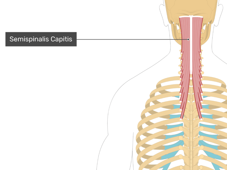
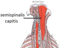
Semispinalis Capitus
Semispinalis Capitus belongs to the semispinalis group of the muscle, which in turn is part of the Transversospinal group of muscles (It is formed of muscles between a spinous process and the transverse process of the vertebrae below).
The semispinalis muscles have the longest fascicles of the transversospinalis group of the muscles, spanning six segments. The muscles in this group are the semispinalis capitis, semispinalis cervicis, and semispinalis thoracis muscles.
The semispinalis capitis is a long slender muscle that provided a long moment arm to provide an efficient extension of the neck.
Origin:
The Semispinalis capitis muscle originates from the articular processes of the C 5, C6, C7, and C8 as well as the transverse processes of T 1, T2, T3, T4, T5, and T6.
Insertion:
The semispinalis capitis muscles insert onto the occiput in between the superior and inferior nuchal lines.
Nerve supply:
The nerve supply of the semispinalis capitis muscle is the greater occipital nerve, which additionally innervates the scalp
Blood supply:
The blood supply of the Semispinalis capitis muscles is descending branches of the occipital artery and the superior intercostal artery, via the dorsal rami of the upper two posterior intercostal arteries
Action:
Acting bilaterally: Extension of the head and neck.
Acting unilaterally: Rotation of the head and neck to the opposite side
Semispinalis Cervicis
Semispinalis Cervicis belongs to the semispinalis group of muscles, which in turn is part of the Transversospinal group of muscles (It is formed of muscles between a spinous process and the vertebrates transverse process below).
The semispinalis muscles have the longest fascicles of the transversospinalis group of the muscles, spanning six segments. The muscles in this group include the semispinalis capitis, semispinalis cervicis, and semispinalis thoracis muscles.
Origin:
The Semispinalis Cervicis muscles originate from the transverse processes of T1 to T6, articular processes of the 4th to 7th cervical vertebrae
Insertion:
The Semispinalis Cervicis muscles insert into the Spinous processes of C2 to C5
Nerve supply:
The nerve supply of the Semispinalis Cervicis muscles is the dorsal rami of cervical spinal nerves.
Blood supply:
The blood supply of the Semispinalis Cervicis muscles is the deep cervical artery
Action:
Acting bilaterally: extension of the cervical spine
Acting unilaterally: lateral flexion of the neck and rotation to the neck’s opposite side.
The Deepest layer of the muscles on the back of the neck
Rectus capitis posterior major muscle
Rectus capitis posterior major muscle is one of four small suboccipital muscles; with rectus capitis posterior minor muscle, obliquus capitis inferior muscle, and obliquus capitis superior muscle being the other three.
When both muscles contract bilaterally they act to the extension of the head on the neck, while the unilateral contraction rotates the head ipsilaterally at the atlantoaxial joint.
Origin:
The Rectus capitis posterior major muscle originates from the Tip of the Spinous process of the axis(C2)
Insertion:
The Rectus capitis posterior major muscle inserted into The lateral part of an inferior nuchal line of the occipital bone
Nerve supply:
The nerve supply of The Rectus capitis posterior major muscle is the Suboccipital nerve (posterior ramus of spinal nerve C1)
Blood supply:
The blood supply of The Rectus capitis posterior major muscle is the Vertebral artery and descending branches of the occipital artery
Action:
Bilateral contraction at the atlantooccipital joint: extension of the Head
Unilateral contraction at the atlantoaxial joint: rotation of the Head (ipsilateral- Same side)
Rectus capitis posterior minor muscle
Rectus capitis posterior minor muscle is a paired muscle that is located in the suboccipital compartment of the neck. It is part of the suboccipital group of the muscle which comprises four muscles in total, the other three being rectus capitis posterior major, obliquus capitis inferior, and obliquus capitis superior muscles.
Origin:
The Rectus capitis posterior minor muscle originates from the tubercle on the posterior arch of the atlas (C1)
Insertion:
The Rectus capitis posterior minor muscle is inserted on the Medial portion of the inferior nuchal line of the occipital bone.
Nerve Supply:
The nerve supply of the Rectus capitis posterior minor muscle is the Suboccipital nerve or dorsal ramus of the cervical spinal nerve (C1)
Blood Supply:
The blood supply of the Rectus capitis posterior minor muscle is the vertebral artery and the deep descending branch of the occipital artery.
Action:
Extension of the head
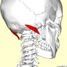
Obliquus capitis superior muscle
Obliquus capitis superior, or superior oblique muscle, is a small paired muscle that is located deep in the upper cervical region, at the base of the occipital bone. It is one of four muscles that comprise the suboccipital group of the muscles along with rectus capitis posterior major, rectus capitis posterior minor, and obliquus capitis inferior muscles.
Obliquus capitis superior muscles contribute to the extension of the head. However, Obliquus capitis superior muscles’ role as a postural muscle is more significant than its role as a prime mover.
Origin:
The Obliquus capitis superior muscle originates from the Superior surface of the transverse process of the atlas (C1)
Insertion:
The Obliquus capitis superior muscle inserted Between the superior and inferior nuchal lines of the occipital bone
Nerve Supply:
The nerve supply of The Obliquus capitis superior muscle is the Suboccipital nerve or dorsal ramus of the cervical spinal nerve (C1)
Blood Supply:
The blood supply of The Obliquus capitis superior muscle is the vertebral artery and the deep descending branch of the occipital artery.
Action:
Bilateral contraction – Atlantooccipital joint: extension of the Head
Unilateral contraction – Atlantoaxial joint: lateral flexion of the head(ipsilateral-Same side)
Obliquus capitis inferior muscle
Obliquus Capitis Inferior muscle (also known as the Inferior Oblique) is a small muscle that runs posteriorly and inferomedially from C1 to C2. Obliquus Capitis Inferior muscle is situated under the deep cervical vein and comprises the inferior border of the suboccipital triangle. Obliquus Capitis Inferior muscle is the only suboccipital muscle that does not attach to the skull.
Obliquus capitis inferior is one of the four suboccipital muscles. Being grouped with rectus capitis posterior major muscle, rectus capitis posterior minor muscle, and obliquus capitis superior muscles; obliquus capitis inferior muscle is the largest muscle of the four.
The functions of obliquus capitis inferior muscle are to facilitate the movements of the head and neck and maintain posture by supporting the atlantoaxial joint.
Origin:
The Obliquus capitis inferior muscle originates from the Base of the spinous process and adjoining lamina of the axis.
Insertion:
The Obliquus capitis inferior muscle inserts Along the inferior aspect of the tip of the transverse process of the atlas.
Nerve Supply:
The nerve supply of The Obliquus capitis inferior muscle is the Suboccipital nerve or dorsal ramus of the cervical spinal nerve (C1).
Blood Supply:
The blood supply of The Obliquus capitis inferior muscle is the Vertebral artery and the deep descending branch of the occipital artery.
Action:
Bilateral contraction – Atlantooccipital joint: extension of the Head
Unilateral contraction – Atlantoaxial joint: rotation of the Head (ipsilateral-Same side)
The Common conditions that affect the neck muscles which include:
- Spasms: Spasm also known as muscle cramps, muscle spasms occur when a muscle contract and can’t relax. Most spasms are short, lasting only for a few seconds. But you might have a sore or stiff neck afterward.
- Strains: A muscle strain is an injury to the muscle or the tendon. It occurs due to overstretching or tearing the muscle fibers.
- Whiplash: If your head suddenly moves forward and then whips backward, you can injure the soft tissue in your neck. Whiplash usually involves the muscles, ligaments, and tendons.
Exercise of the muscle of the back of the neck
Chin tuck
Sit straight and look straight ahead with the ears directly over the shoulders.
Place a finger on the chin.
Without moving the finger, pull the chin and head straight backward until a good stretch is felt at the base of the head and top of the neck.
Hold for 5 seconds if possible.
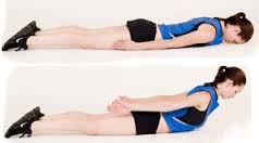
Prone Cobra
This workout is done lying face down on the ground and uses gravity as resistance in the reinforcing process.
Lying face down, put the forehead on a rolled-up hand towel for comfort.
Place the arms at the side, palms down on the ground.
Place your tongue on the roof of your mouth
Pinch the shoulder blades together and lift the hands off the ground.
Roll the elbows in, palms out, and thumbs upward.
Gently lift the forehead about an inch off the towel keeping the eyes looking straight at the ground
Hold the position for 10 seconds.
Perform 10 repetitions.
Stretching exercise
Splenius capitis muscle stretch
The left-side splenius capitis is stretched by flexing, right laterally flexing, and right rotating the head and neck at the spinal joints, while the left-side shoulder girdle is allowed to elevate.
Splenius cervicis muscle stretch
Stand or sit upright
Keep your head up facing straight ahead
Push your head forward side by sticking out your chin
Note: Keep your head up during this stretch. Do not let your chin fall towards the floor.
Trapezius muscle stretch
Slowly take your left ear toward your left shoulder. It’s natural for your right shoulder to lift as you do this. If that happens, ease your head back toward the center until you can relax your right shoulder back down.
Gently release this side, and then ease your right ear toward your right shoulder and complete the stretch on the other side, breathing deeply through it.
You can hold for 30 seconds and 3 repetition

Semispinalis capitis muscle stretch
The semispinalis capitis muscle(of the transversospinalis group) is stretched by flexing the head and neck at the spinal joints. Adding in lateral flexion (not seen in the accompanying illustration) will increase the efficacy of the stretch for the opposite-side semispinalis capitis muscle.
Splenius cervicis muscle stretch
The left-side splenius cervicis muscle is stretched by flexing, right laterally flexing, and right rotating the neck at the spinal joints, while the left-side shoulder girdle is allowed to elevate.
Suboccipital Muscle Stretch
Manual Stretching
Patient position and procedure: Sitting
Identify the spinous process of the 2nd cervical vertebra and stabilize it with your thumb or with the second metacarpophalangeal joint (and the thumb and index finger around the transverse processes). Asked the patient slowly
nodding, doing only a tipping motion of the head on the upper spine. Guide the movement by placing the other hand across the forehead of the patient.
Patient position and procedure: Supine
Sit on a stool at the head of the treatment table with your forearms resting on the table. One hand stabilizes the C2 vertebra by grasping the transverse processes between the proximal part of the thumb and index finger; the other hand supports the occiput. Nod the head of the patient with the hand under the occiput to take up the slack of the suboccipital muscles; then ask the patient to roll the eyes upward. This causes a gentle isometric contraction of the muscles of the suboccipital. After holding seconds 6, ask the patient to roll the eyes downward. As the suboccipital muscles relax, take up the slack by passively nodding the head through a new range. The only motion should occur between the occiput and C2. The contraction is gentle in order to not cause overflow into the multisegmental erector spinae muscles and upper trapezius muscles.
Self-Stretching
Patient position and procedure: Supine or sitting. Instruct the patient to nod the head, bringing the chin toward the larynx until a stretch is felt in the suboccipital region. Putting light pressure under the occipital area with the palm of your hand while tipping the patient’s head forward reinforces the motion.