Obturator Externus Muscle: Origin, Insertion, Function, Exercise
Table of Contents
Obturator Externus Muscle Anatomy
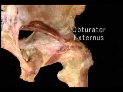
Obturator externus muscle is a flat, triangular-shaped, paired muscle located in the gluteal region. It is situated on the anterior side of the obturator foramen, attached to the obturator membrane and the related margin of the obturator foramen.
The muscles play an important role in the gluteal region, The muscle helps to external rotation of the femur when the hip is extended but when the hip is flexed, it adducts the femur. It also provides stability to the hip joint.
In this article, we discuss the Anatomy, function, and exercise of the muscles.
Obturator externus origin:
Outer obturator membrane, the rim of pubis, and ischium bordering it (obturator foramen).
Insertion:
Trochanteric fossa on the medial surface of the greater trochanter.
Nerve supply:
The posterior division of the obturator nerve (L2,3,4) of the Lumbar plexus.
Blood supply:
Obturator and medial circumflex femoral arteries.
Action:
- External rotation of the Hip
- Provides stability of the Hip with other associated muscles
- Adduction of the thigh when Hip is flexed


Where is the obturator externus muscle located?
Relation of the muscles:
Obturator externus is located in the pelvis on the anterior aspect of the obturator foramen, attached to the obturator membrane and the related margin of the obturator foramen. The obturator foramen is enclosed by the muscles and deep to the pectineus and superior parts of the adductors of the thigh.
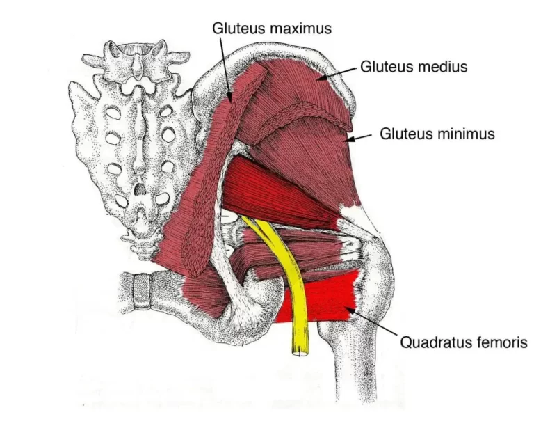
Obturator externus muscle-tendon is located deep to the quadratus femoris muscle and it also separates from the neck of the femur.
The obturator vessels mainly anterior and posterior branches of the obturator artery and vein are located deep to the obturator externus muscle, on the outer surface of the obturator membrane. There are also obturator nerves traveling in close proximity to this muscle. The anterior branch of the nerve passes over the anterior surface of the muscle while the posterior branch crosses the muscle before both branches descend to innervate the muscles of the thigh.
Obturator externus Exercise:
There are mainly two types of exercise: strengthening exercise and stretching exercise.
Lateral rotators stretching exercise:
To do this exercise, sit in a chair and maintain an erect posture. Start with placing the left ankle over your right thigh. Hold the left knee with your both hands and pull it toward the right shoulder. Exercise must be a pain-free gentle stretch. Hold this for 10-20 seconds and relax. Repeat on another leg. Do this exercise 3 to 4 times a week.
Hip adductor stretch
You can take a kneeling position on a soft mat with your left foot while your right foot is in front of you, while your right foot is ninety degrees. Gradually slide the left knee to the side, keep the chest up, hold for 10 to 20 seconds, and relax. Repeat on another leg. Do this exercise 3 to 4 times a week.
Clinical importance
Obturator externus muscle tear is a rare overuse sports injury due to mainly Repetitive eccentric contraction of muscles. Symptoms mainly in the groin area and radiated to the buttock area. Pain and tenderness at the ischial tuberosity. on palpation is also seen.
Treatment mainly uses of RICE Principle and after 2 to 3 weeks gradual pain-free strengthening and stretching exercises help to return to the sports.
Using a posterior approach, Solomon et al. investigated the function of the short external rotators in hip stability after total hip replacement (THR). They also out that maintaining the external obturator and piriformis lowers the chance of dislocation following total hip replacement, suggesting alternative methods. OE stabilizes the hip joint by reinforcing the posterior capsule with its fibers.
The bursa of the obturator externus (OE) muscle was located between the transverse acetabular ligament and the OE muscle. It contained bursal fluid. Research indicates that OE bursa is more common in intra-articularly pathological hips than in healthy hips.
In professional basketball players, repetitive eccentric contraction of the obturator externus results in musculotendinous tears. A quick and trouble-free return to competition is guaranteed by targeted rehabilitation.
Impingement syndrome following total hip replacement: If the transverse acetabular ligament is released or if the acetabular cup position protrudes past the caudal rim following arthroplasy, there is a strong correlation between the musculo-tendinous portion of the OE muscle and the inferior margin of the acetabulum. The chance of impingement was reduced by releasing the OE insertion connection from the posterior capsule.
FAQ
The primary function of the obturator externus muscle is to rotate the thigh laterally at the hip joint. It also contributes to the general mobility and control of the lower limb and helps to stabilize the hip joint.
No, it is difficult to palpate the obturator externus muscle directly because it is located deep within the pelvis.
The front rims of the pubis and ischium, as well as the outside of the obturator membrane, are the origins of the obturator externus. It attaches to the greater trochanter’s posterior side of the femur. The rounded region on the upper, outside portion of the femur is called the greater trochanter.
The ipsilateral base moves posteriorly and superiorly as a result of the piriformis pulling the sacral apex anteriorly and laterally. The ilia crest moves anteriorly as a result of the obturator externus pulling the lower pelvis posteriorly.
The fibers entered the trochanteric fossa as a cylindrical tendon, with some of the fibers continuing towards the piriformis fossa. They passed laterally along the inferior margin of the acetabulum, functioning as a sling at the inferior portion of the neck.
The flat, triangular external obturator muscle, also known as the obturator externus muscle, covers the exterior of the pelvic anterior wall. It is alternately regarded as a component of the gluteal area and the medial compartment of the thigh.
The posterior branch of the obturator nerve (L3 and L4), which originates in the lumbar plexus, innervates the obturator externus.
Professional male soccer players may experience acute onset groin and anterior hip pain following long-distance passing or shooting due to an obturator externus tear. A comparatively quick healing period and conservative care are made possible by accurately diagnosing obturator externus injuries.

