Temporalis muscle
| | |

Temporalis Muscle Anatomy

Introduction of the Temporalis muscle In anatomy, the temporalis muscle is also known as the temporal muscle. This muscle fills the temporal fossa. The temporalis muscle is a thin, fan-shaped muscle located within the temporal fossa of the skull. Along with the medial pterygoid muscle, lateral pterygoid, and masseter muscles, it belongs to the group…

Masseter muscle
| | |

Masseter Muscle Anatomy

Introduction The masseter is a quadrilateral muscle that covers the lateral surface of the ramus of the mandible. The masseter muscle fibres are arranged in three layers. The masseter muscle is a paired, strong, thick, and rectangular muscle that originates from the zygomatic arch and extends down to the mandibular angle. It contains a superficial…

Medial pterygoid muscle
| | |

Medial pterygoid muscle Anatomy

Introduction The medial pterygoid is a thick, quadrilateral muscle of the face and it has a small superficial head and a large deep head which forms the major part of the muscle.The medial pterygoid muscle is supplied by the mandibular branch of the trigeminal nerve (V). It is important in mastication (chewing). It belongs to…

The semispinalis thoracis
| |

Semispinalis Cervicis

Introduction Semispinalis Cervicis belongs to the semispinalis group of the muscle, which in turn is part of the Transversospinal group of muscles (It is formed of muscles between a spinous process and the vertebrates transverse process below). The semispinalis muscles have the longest fascicles of the transversospinalis group of the muscles, spanning six segments. The…

The semispinalis thoracis
|

Semispinalis Thoracis

Introduction The semispinalis thoracis muscle contains thin, narrow, fleshy fasciculi, interposed between tendons of considerable length. Semispinalis thoracis muscle consists of five fascicles bridging over five to six vertebral levels between the transverse and spinous processes of certain cervical vertebrae and thoracic vertebrae. With these attachments, semispinalis thoracis muscle aids several movements of the vertebral…

Popliteus muscle
| |

Popliteus Muscle Anatomy

Introduction the popliteus muscle is a major stabilizer of the knee jointThe popliteus is a small, thin, flat, triangular-shaped musculotendinous complex muscle that forms the floor of the popliteal fossa. It belongs to the deep posterior leg muscles, along with the tibialis posterior, flexor digitorum longus, and flexor hallucis longus muscles. It is a deep…

The diaphragm
| |

Diaphragm Muscle

Introduction The diaphragm is an unpaired, dome-shaped skeletal muscle that is located inside the trunk. It is the primary muscle that is active in inspiration. Contraction of the diaphragm muscle facilitates the expansion of the thoracic cavity. This increases the volume of the cavity, which in turn reduced the intrathoracic pressure allowing the lungs to…

Peroneal muscles
| |

The peroneal muscles

Introduction The fibular/peroneal muscles are the two muscles of the lateral compartment of the leg. These muscles are the fibularis longus and fibularis brevis. The peroneal muscles Originate from the fibula and are inserted onto the plantar surfaces of certain tarsal and metatarsal bones, these muscles play an important role in the movements of the…

Muscle of the medial side of thigh
| | |

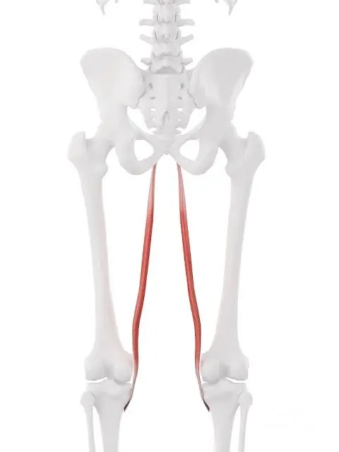
Muscle of medial compartment of the thigh

Introduction The hip adductors are a group of muscles located in the medial compartment of the thigh. These muscles include the adductor longus, adductor brevis, adductor Magnus, gracilis, and pectineus. Due to their position, the hip adductors shape the surface anatomy of the medial side thigh. More specifically, these muscles extend from the anteroinferior external…