Spinalis Cervicis
| |

Spinalis Cervicis

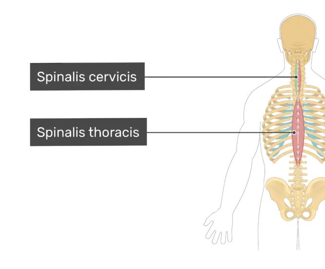
Introduction of the Spinalis Cervicis The Spinalis group of the muscle is part of the erector spinae (ES) group (the intermediate layer of the intrinsic back muscles). Spinalis Cervicis muscle is the cervical portion of the spinalis muscle with spinalis capitis muscle superiorly and spinalis thoracis muscle inferiorly.The Spinalis Cervicis muscle is variably present. Origin:…

| |

Suprahyoid muscles

Introduction of the suprahyoid muscles The suprahyoid muscles are the digastric, the stylohyoid, the mylohyoid, and the geniohyoid. They all are pharyngeal muscles, with the exception of the geniohyoid muscle.They are described below. The digastric muscle The digastric muscle lies below the body of the mandible, and extends, in a curved form, from the mastoid…

Mylohyoid muscle
| |

Mylohyoid muscle

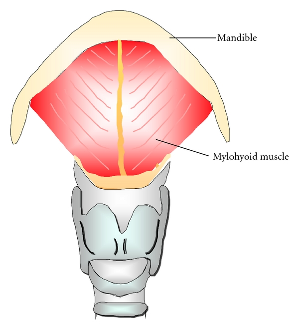
Introduction The mylohyoid muscle is one of the suprahyoid muscles.The mylohyoid muscle is a flat, triangular muscle lying deep in the anterior belly of the digastric. Both the sides of the mylohyoid muscles together form the floor of the mouth. Along with the other suprahyoid muscles (the digastric, the geniohyoid, and the stylohyoid), The mylohyoid…

Lateral pterygoid Muscle
| | |

Lateral pterygoid Muscle

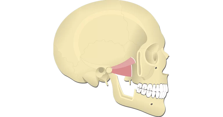
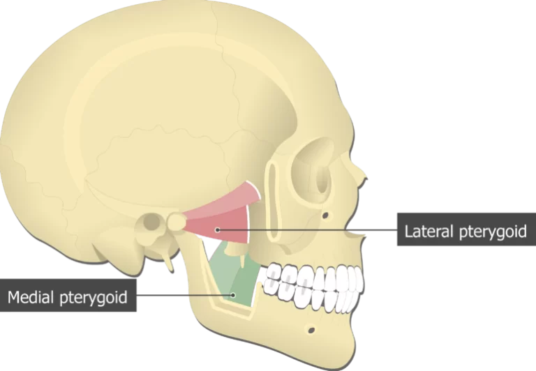
Introduction The lateral pterygoid muscle has two heads the upper head and the lower head.The lateral pterygoid is the fan-shaped muscle located laterally in the infratemporal fossa of the skull. It is one of the four mastication muscles, along with the medial pterygoid muscle, temporalis, and masseter muscles. All these mastication muscles act upon the…

Temporalis Muscle Anatomy
| | |

Temporalis Muscle Anatomy

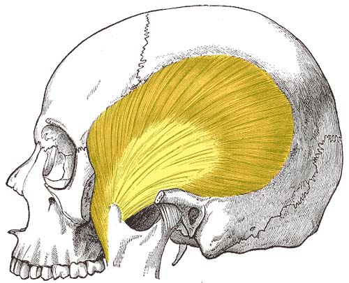
Introduction of the Temporalis muscle In anatomy, the temporalis muscle is also known as the temporal muscle. This muscle fills the temporal fossa. The temporalis muscle is a thin, fan-shaped muscle located within the temporal fossa of the skull. Along with the medial pterygoid muscle, lateral pterygoid, and masseter muscles, it belongs to the group…

The masseter muscle Anatomy
| | |

The masseter muscle Anatomy

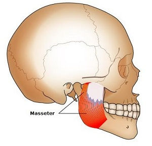
Introduction The masseter is a quadrilateral muscle that covers the lateral surface of the ramus of the mandible. The masseter muscle fibres are arranged in three layers. The masseter muscle is a paired, strong, thick, and rectangular muscle that is originating from the zygomatic arch and extends down to the mandibular angle. It contains a…

Medial pterygoid muscle Anatomy
| | |

Medial pterygoid muscle Anatomy

Introduction The medial pterygoid is a thick, quadrilateral muscle of the face and it has a small superficial head and a large deep head which forms the major part of the muscle.The medial pterygoid muscle is supplied by the mandibular branch of the trigeminal nerve (V). It is important in mastication (chewing). It belongs to…

Semispinalis Cervicis
| |

Semispinalis Cervicis

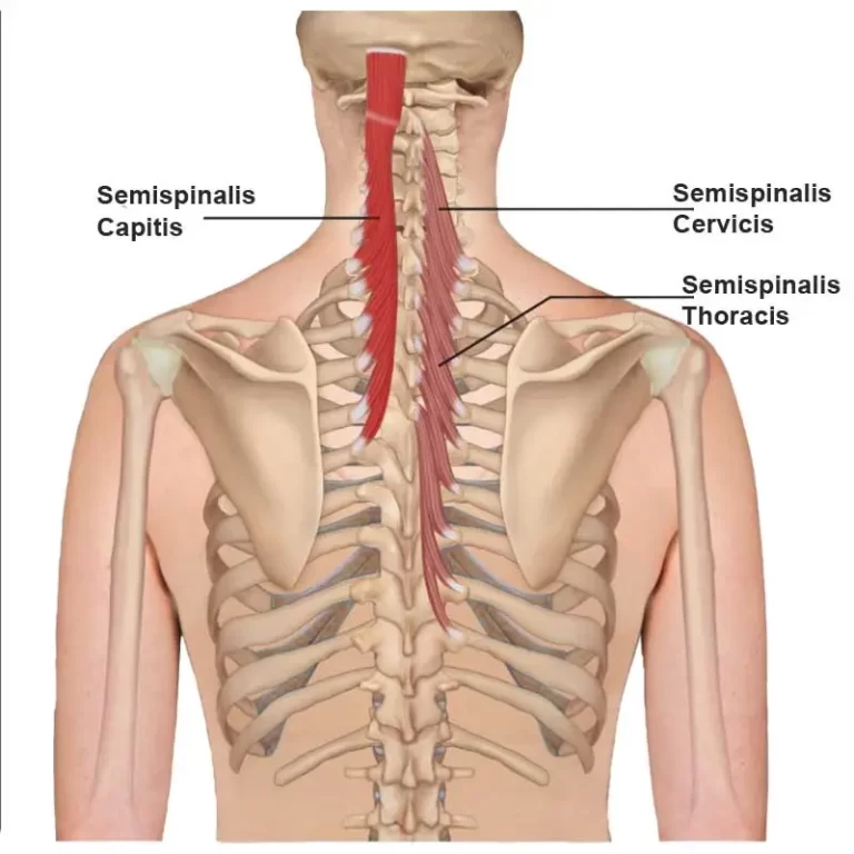
Introduction Semispinalis Cervicis belongs to the semispinalis group of the muscle, which in turn is part of the Transversospinal group of muscles (It is formed of muscles between a spinous process and the vertebrates transverse process below). The semispinalis muscles have the longest fascicles of the transversospinalis group of the muscles, spanning six segments. The…