Popliteus Muscle Anatomy
Table of Contents
Introduction
the popliteus muscle is a major stabilizer of the knee joint
The popliteus is a small, thin, flat, triangular-shaped musculotendinous complex muscle that forms the floor of the popliteal fossa. It belongs to the deep posterior leg muscles, along with the tibialis posterior, flexor digitorum longus, and flexor hallucis longus muscles.
It is a deep muscle of the knee joint, making the floor of the popliteus fossa. It also makes the lateral musculature of the knee joint, along with the iliotibial band.
It is one of the main posterolateral stabilizers of the knee joint, causing both medial rotation and lateral rotation of the knee, thereby being involved in both the closed-chain phase and open-chain phase of the gait cycle. It also helps stabilizer in regard to internal rotation, anterior translation, and varus force.
Origin of popliteus muscle:
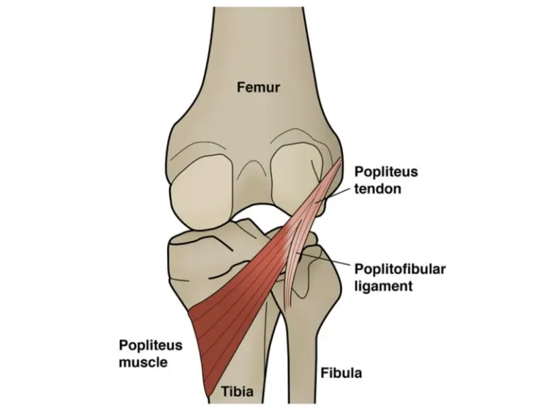
Popliteus muscles originate from, The lateral surface of the lateral condyle of the femur bone,
Origin is intracapsular And the Outer margin of the lateral meniscus of the knee joint
Insertion of popliteus muscle:
The Popliteus muscle inserted on the posterior surface of the shaft of the tibia above the soleal line
Nerve supply:
The nerve supply of the popliteus muscles is the tibial nerve (L5 to S2).
From spinal nerve roots L5 through S1, the tibial nerve innervates the popliteus. There are roughly two to three parallel tibial nerve branches, according to earlier research. The lateral distal border of the muscle located inferior to the fibular head is where the nerve enters the body. The nerve divides into anterior, medial, and lateral distributions within the muscle after entering.
Blood supply:
The popliteus receives blood supply mainly from branches of the popliteal artery, namely the inferior medial genicular arteries and lateral genicular arteries.
Lymphatic Drainage
The popliteal nodes and the deep inguinal nodes of the thigh are the destinations of lymphatic drainage. It is crucial to map and measure lymphatic flow when thinking about infectious or metastatic processes.
Action of popliteus muscle:
Unlocks knee joint by External rotation of the femur on the tibia to flexion.
Muscle Relations
The popliteus is located in the deep posterior compartment of the leg, one of the leg’s four compartments. Next to the popliteus in the deep posterior compartment are the following muscles:
Flexor hallucis longus: Inserts into the base of the great toe’s distal phalanx after originating at the distal part of the fibula and inferior interosseous membrane. In addition, contraction acts as a supplementary plantar flexor and flexes the great toe.
Flexor digitorum longus: The broad tendon of the fibula and the posteromedial side of the tibia are the origins of the flexor digitorum longus muscle. In addition to receiving innervation from the tibial nerve, insertion is visible at the distal phalanges of digits 2 through 5. (S1, S2). It allows for the flexion of toes two through four, plantarflexion of the ankle and support for longitudinal arches.
Tibialis posterior: The posterior fibular surface, the posterior tibial surface inferior to the soleal line, and the interosseous membrane are the sources of the tibialis posterior. The bases of metatarsals 2 to 4 as well as the tuberosity of the navicular, cuneiform, cuboid, and sustentaculum tali of the calcaneus are where the tibialis posterior enters. The tibial nerve innervates it. This muscle is a key ankle inverter and plantar flexor.
Anatomical Variations
The popliteal tendon may occasionally have a little sesamoid bone known as a cyamella submerged in it. Sesamoid bones are auxiliary bones that support physiologic function and are usually found in muscles or tendons. The cyamella can be found at the musculotendinous junction, which is situated superior to the joint line, or in the tendon inferior to the joint line. The lateral condyle of the tibia is where articulation is normally found, surrounding the fibular head. This variation can induce pain similar to a posterolateral meniscal rupture, although it usually does not become physiologically crippling.
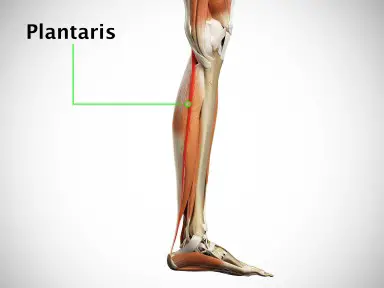
Compared to the popliteus, the peroneal tibialis and popliteus minor muscles are rarely observed. If it exists, the popliteus minor attaches to the knee’s posterior ligament and is located medial to the plantaris. A small percentage of the general population also exhibits the peroneal tibialis. If it exists, it will be located medial to the fibular head, below the popliteus, and extending to the superior part of the tibia’s oblique line. There hasn’t been much discussion of either structure in the recent literature.
Clinical significance
Trauma, poor movement patterns, and posture can often weigh heavily on the popliteus muscle leaving it prone to weakness and getting injured
Popliteus strain: Popliteus muscle injuries are rarely isolated injuries and are most commonly associated with other injuries of the posterolateral corner of the knee such as lateral collateral ligament injury, Anterior cruciate ligament injury, posterior cruciate ligament injury, or meniscal injury.
Popliteal tendinopathy can also occur as posterolateral pain in the knee. However, it can be difficult to single it out due to other more common knee pain causes in the vicinity. As this muscle inhibits excessive rotation of the tibia along with preventing significant anterior translation of the knee, it can be pathologically overcome secondary to excessive sprinting or running downhill, and hence such types of activities should be avoided or modified to run on flat surfaces like a treadmill.
Physiotherapy treatment for Popliteal muscle
Popliteal muscle release
Long sitting with a lacrosse ball behind band knee and searching for tender point areas. Slowly add pressure. If tender or numb, move to a slightly different area, and add movement by internal rotation and external rotation of the lower leg.
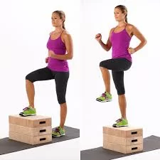
Step up and step down exercise
Place one foot on top of a small step stool. Keeping the raised leg slightly bent at the knee, step forward with the opposite leg. Next, step backward, then to the right and left of the foot planted on the step. Repeat this movement for 20 to 30 repetitions.
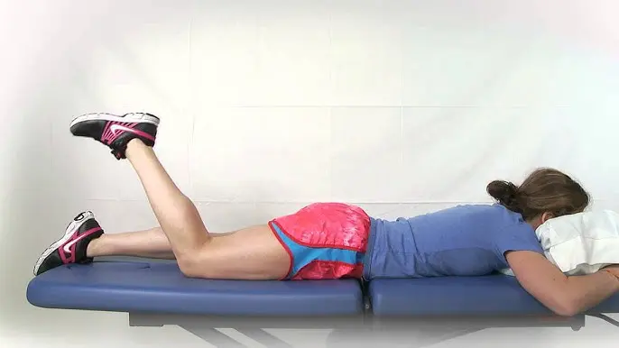
Prone hamstring curl
Lie down on your stomach
Bend your knee to pull your heel toward your buttock, keeping your thighs and hips on the floor.
Stop when you can’t pull any further. Return to starting position.
Do 10 to 15 repetitions
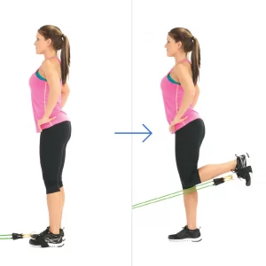
Exercise with a resistance band
Wrap a resistance band around your ankle
The foot on the non-weight-bearing limb moves behind the stance leg via external rotation of the hip and knee flexion.
The foot of the non-weight-bearing limb continues to move behind the stance leg with increasing internal tibial rotation.
Popliteus muscle stretch in long sitting
Sit straight on the ground with your legs straight out in front of you.
If it helps, bend the leg you do not plan to stretch and place it where it helps to support you.
Loop a strap or a band around the ball of your other foot.
We can also use a thick resistance band.
Ensure your knee and toes point up directly towards the ceiling.
Gather any excess
Sitting up straight, tighten your thigh muscles as you pull back on the band.
Allow your heel to stretch away from you. Hold this position for 20 to 30 seconds and then relax. Repeat 3 times.

2 Comments