Subcostalis Muscle
Table of Contents
Subcostalis Muscle Anatomy
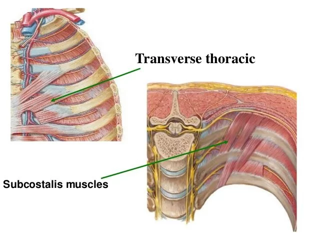
Subcostalis muscle are the narrow muscles that span two or three intercostal gaps and are located on the inner surface of the posterior thoracic wall. They make up the intrinsic musculature of the chest wall along with the intercostal, serratus posterior, levatores costarum, and transversus thoracis muscles.
The subcostal muscles maintain the thoracic cage and intercostal gaps as well as aid in respiration by pulling the ribs inferiorly during forced exhale.
The subcostal muscle’ anatomy and function will be covered in this article.
Subcostalis muscle consist of muscular and aponeurotic fasciculi.
Origin:
inner surface of one rib.
Insertion:
inner surface of the second or third rib below, near its angle.
Nerve supply:
intercostal nerves.
Blood supply:
Posterior intercostal artery, musculophrenic artery
Action:
The function of the subcostalis muscles is to assist breathing by Depresses lower ribs during forced exhalation, as well as to maintain the intercostal spaces and thoracic cage.
The purpose of Subcostalis muscle, which are part of the accessory respiratory musculature, is to depress the ribs during forced expiration. They can do this by pulling the ribs inward towards the axis of the thorax, which causes the lungs to be compressed and expel air.
Relations
The thoracic vertebrae’s lateral border is where subcostal muscles are located. The innermost intercostal muscles’ lateral borders come into contact with them and may occasionally merge with them. The internal intercostal membranes and the inside surface of the ribs are covered by their posterior surfaces. These muscles’ anterior surfaces are connected to the parietal pleura and endothoracic fascia.
FAQs
Due to their role in depressing the ribs during forced expiration, subcostalis muscles are considered secondary respiratory muscles. They can do this by pulling the ribs inward towards the axis of the thorax, which causes the lungs to be compressed and expel air.
Posterior chest
The Subcostalis muscle, which belongs to the intercostal muscular group, has a varied architecture. It is located in the posterior chest, close to the rib angles, on the deep surface of the innermost intercostal muscle, typically spanning two to three intercostal gaps.
A similar term for the subcostalis muscle is the “Subcostal muscle and aponeurotic fasciculi”. The same muscle in the human body is referred to by both of these titles. The subcostal muscle is a member of the intercostal muscle group and is situated in the back. The word “subcostal” (sub- = below, costal = ribs) refers to its location slightly below the ribs. The subcostal muscle helps in respiration as its primary function.
The upper (1-4) and lower (9-11) intercostal gaps are where it is most common. According to studies, each person has an average of five Subcostalis muscles, which can either be shaped like a band or a fan.

