Brachial Artery
Table of Contents
Introduction
The brachial artery is a major blood vessel of the upper arm, extending from the shoulder to the elbow. It is the main artery supplying blood to the arm and plays a crucial role in maintaining adequate blood flow to the tissues of the upper limb.
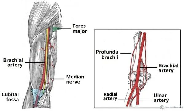
It is the continuation of the axillary artery beyond the lower border of the teres major muscle. It travels along the arm’s ventral surface to the cubital fossa at the elbow. The radial and ulnar arteries, which travel down the forearm, are where it divides after that.
In certain individuals, the radial and ulnar arteries pass through the upper arm, causing the bifurcation to occur much earlier. The brachial artery pulse can be felt on the anterior aspect of the elbow, medial to the biceps tendon. A stethoscope and sphygmomanometer, or blood pressure cuff, are frequently used to measure the pulse rate.
In proximal regions, the median nerve is situated directly lateral to the brachial artery, indicating a strong connection between the two anatomical structures. The median nerve is located anterior to the elbow joint and crosses the medial side of the brachial artery distally.
Because the brachial artery is located close to the skin’s surface, it can be harmed by traumatic injuries such as arm fractures.
What is the brachial artery?
The aortic and brachiocephalic trunks are the respective origins of the left and right subclavian arteries. They go via the shoulder area and are known as the axillary arteries. When the blood vessels reach the arm, they are referred to as the brachial arteries. Your body’s brachial arteries are not connected to one another.
Aortic branches carry blood that has been oxygenated to every part of the body. This function is carried out for the upper limbs via the brachial arteries.
If you’ve ever had your blood pressure checked, the medical professional will wrap an arm cuff over you. Your brachial artery is used by that cuff to assess the pressure inside your arteries. Mostly a Doctor will press on this artery to take your brachial pulse during examination.
The triceps brachii muscle is found in the posterior region of the arm, while the coracobrachialis, biceps brachii, and brachialis muscles are found in the anterior region.
The main artery supplying the arm, the brachial artery, is located inside the anterior compartment. Because of its close relationship to the humerus, it can be used for a variety of clinical exams, including blood pressure and pulse checks, however, it is also susceptible to trauma that primarily affects the bone, such as fractures.
Your doctor may periodically take blood pressure measurements in your lower limbs and arms. The ankle-brachial index is the ratio of the pressures measured at these two sites. It aids in the diagnosis of peripheral artery disease. Blood pressure testing can be used to identify coarctation of the aorta, a congenital heart condition that can cause a child’s upper and lower limb blood pressure to differ.
A further use for the brachial artery is in interventional radiology techniques.Doctors place a small, flexible catheter through the elbow artery and then push it into the major vessels near the heart. This allows them to detect and treat conditions such as blood clots, blockages, and aneurysms in the main blood channels, sometimes avoiding the need for invasive heart surgery.
Anatomy
The major artery in the arm is the brachial artery. It is the axillary artery’s continuation.
The brachial artery is the principal blood vessel supplying the arm, hand, and forearm. It supplies the tissues of the upper limb with nourishment and oxygen.
The ulnar and radial arteries, two terminal branches of the brachial artery, supply blood to the forearm and hand.
The brachial artery is often wounded because of its proximity to the skin. However, since the health of your arm and hand depends on the brachial artery functioning properly, such injuries should always be prevented.
Course:
One of the primary blood vessels supplying the arm and hand is the brachial artery, along with the axillary and subclavian arteries. It is also an essential component of the heart system. It is the continuation of the axillary and subclavian arteries and is situated in the arm between the elbow and the teres major muscle.
The brachial artery descends the underside of the arm after continuing from the axillary artery at the shoulder. It forms the cubital fossa, the triangular pit on the inside of the elbow, together with the bicep tendon and medial cubital vein.
The axillary artery, which passes through the upper chest and armpit, continues as the brachial artery. From your shoulder to the cubital fossa in front of your elbow, the brachial artery travels down your arm. The ulnar and radial arteries are the two branches that emerge from the brachial artery at this point.
The interior of your upper arm is home to the brachial artery. It travels toward the center of your elbow when it gets to the lower portion of your arm. It loops here in front of the elbow joint, between the inner side of the median nerve and the tendon of the biceps muscle on the outer side.
The median nerve, the main nerve supplying your forearm, runs parallel to the brachial artery.
What material makes up the brachial artery?
There are three layers to each artery in your body:
- Tunica intima: This inner layer allows your blood to circulate freely. It keeps pollutants out of your circulation, controls blood pressure, and avoids blood clots.
- Media: The layer in the centre helps blood arteries dilate and constrict so that blood flows in a single direction.
- Adventitia: The outer layer of blood vessels provides stability and structure. Its small capillaries remove waste from your cells and deliver nutrients and oxygen to them.
Structure:
The following branches originate from the brachial artery. Top to bottom, they consist of:
- Profunda brachii artery
- Superior ulnar collateral artery
- Middle ulnar collateral artery (M)
- Inferior ulnar collateral artery
- Radial artery
- Ulnar artery
- Deltoid artery (D)
- Nutrient branches to the humerus
It also results in the development of significant anastomotic networks in the shoulder and elbow (as the axillary artery).
The brachial artery is lateral to the biceps head. For the majority of its length, the median nerve runs medial to the brachial artery.
Profunda Brachi Artery: The large posteromedial branch of the brachial artery, distal to the teres major muscle, is called the profunda brachii artery. It travels beside the radial nerve. The profunda brachii artery passes posteriorly between the long and medial heads of the triceps brachii muscle before entering the humerus through the spiral groove. Subsequently, it bifurcates into two branches: the anteriorly descending radial collateral and the posteriorly descending middle collateral.
Collateral branches: The bigger of the two collateral branches, the middle one, emerges behind the humerus and moves behind the lateral intermuscular septum to the elbow. This branch is located in close proximity to the lateral heads of the triceps brachii and the brachialis. The profunda brachii artery passes posteriorly between the long and medial heads of the triceps brachii muscle before entering the humerus through the spiral groove.
Subsequently, it bifurcates into two branches: the anteriorly descending radial collateral and the posteriorly descending middle collateral. It reaches the brachioradialis in back and the lateral head of the triceps brachii in front. The interosseus recurrent artery is reached posterior to the lateral epicondyle by the middle collateral branch, which either stays deep into the fascia or crosses it to become cutaneous. Five fasciocutaneous perforators are released, small arteries that pierce the fascia, separate muscles, and supply blood to the skin.
Radial Collateral branch: The radial collateral branch runs parallel to the radial nerve and crosses the lateral intermuscular septum before descending anteriorly to the lateral epicondyle between the brachialis and brachioradialis. The radial nerve, the brachioradialis and brachialis muscles, and a number of fasciocutaneous perforators are supplied by it before it joins the radial recurrent artery.
Nutrition artery: The nutrition canal, which is essentially a sizable foramen or hole in the humerus, is where the nutrient artery enters after emerging from the brachial artery around the middle level of the arm. It passes posterior to the deltoid tuberosity and into this canal near the coracobrachialis connection.
Superior ulnar collateral artery: The superior ulnar collateral artery begins slightly distal to the arm’s mid-level and arises from the brachial artery, though it can also infrequently emerge as a branch of the profunda brachii artery. It travels down the ulnar nerve, passing through the posterior compartment of the arm and the medial head of the triceps brachii muscle.
It then moves between the humerus’s medial epicondyle and the ulnar epicondyle. It joins the inferior collateral artery deep within the flexor carpi ulnaris muscle to the posterior branch of the posterior ulnar recurrent artery. Occasionally, a branch will travel anteriorly to the medial epicondyle before joining the anterior ulnar recurrent artery in anastomosis.
Middle ulnar collateral artery: The middle ulnar collateral artery, which in some people supplies the triceps brachii muscle, is derived from the brachial artery, which is located between the superior and inferior ulnar collateral arteries. The anterior branch of the anterior ulnar recurrent artery is where it anastomoses after moving anteriorly to the medial epicondyle. Similar to the middle collateral branch of the profunda brachii artery, it releases a few small fasciocutaneous perforators.
Inferior ulnar collateral artery: The inferior ulnar collateral (supratrochlear) artery originates from the brachial artery and extends about 5 centimeters forward of the elbow joint. It crosses medially between the brachialis muscle and the median nerve before crossing the medial intermuscular septum.
It then forms an arch proximally to the olecranon fossa, a depression on the posterior face of the humerus, by wrapping around the humerus’s triceps brachii muscle and bone in a spiral pattern until it anastomoses with the profunda brachii artery’s middle collateral branch.. The inferior ulnar collateral artery splits anteriorly to the brachialis muscle, forming a few branches that either flow posteriorly to the posterior ulnar recurrent artery and superior ulnar collateral artery or anteriorly to the medial epicondyle, where they anastomose with the anterior ulnar recurrent artery.
Deltoid artery: The deltoid artery is a muscular branch of the brachial artery that extends between the lateral and long heads of the triceps brachii. It continues until it joins the descending branch of the posterior humeral circumflex artery.
Radial artery: The radial artery descends deeply to the brachioradialis in order to nourish the radial recurrent artery distal to the elbow joint.. It passes between the superficial and deep branches of the radial nerve before continuing superiorly posteriorly to the brachioradialis muscle and anteriorly to the supinator and brachialis muscles. Before anastomosing with the radial collateral branch of the profunda brachii artery, it supplies the brachialis, supinator, brachioradialis, and elbow joint.
Ulnar artery: The ulnar artery is the largest terminal branch of the brachial artery. Its initial branch, the ulnar recurrent artery, divides into anterior and posterior branches subsequently. The common interosseous artery, dorsal, palmar, and deep carpal branches, as well as the superficial palmar arch, are all produced by the ulnar artery, which descends the forearm and contributes to the radial artery’s branches and the blood flow to the hand and forearm.
Function:
Blood rich in oxygen is mostly sent to the arm and hand via the brachial artery.
- Biceps muscle
- Muscles called brachialis (behind your biceps).
- Elbow joint.
- The triceps are also known as the brachii muscles.
The blood’s oxygen and nutrients are essential for the healthy function and healing of your arm’s bones, soft tissues, and nerves. The brachial artery, like every other artery in your body, drains oxygen-rich blood from your heart.
The brachial artery is used by clinicians to assess blood pressure since it is somewhat below skin level, particularly around the elbow. This explains why the inflated cuff of a conventional blood pressure gauge is placed on the elbow.
Surgeons may also need to perform brachial artery compression in trauma patients in order to minimize blood loss. This is done proximally above the site of injury to stop blood flow until the patient gets to the operating room. Tissue loss is more likely the longer a tourniquet is inflated.
Tests for brachial artery:
The brachial artery is a crucial point of entry for interventional radiology procedures. Your healthcare provider may implant a catheter—a tiny, flexible tube—into your brachial artery. They thread it up to the blood vessels close to your heart using imaging guidance. Using this minimally invasive technique, your provider can treat issues such as blood clots, aneurysms, or restricted arteries without requiring significant open heart surgery.
Your doctor may also use the ankle-brachial index test (ABI). Your brachial and ankle arteries’ blood pressures are compared throughout this examination. You may have peripheral artery disease (PAD) if your legs aren’t getting enough blood flow.
Anatomical Relationships of the Brachial Artery:
In clinical practice, the brachial artery’s relationship to other arm structures might be significant. The brachial artery is a superficial vessel that is only covered by the skin’s layers and the superficial and deep fasciae, with a few notable exceptions.
The biceps brachii muscle’s bicipital aponeurosis, which covers the artery and divides it from the median cubital vein, is the first location where this is not the case. This occurs at the cubital fossa. The second exception happens when the median nerve travels via the brachial artery near the distal end of the coracobrachialis.
The profunda brachii artery and the radial nerve divide the brachial artery posteriorly from the long head of the triceps brachii muscle. The medial head of the triceps brachii muscle and the attachments of the coracobrachialis and brachialis muscles are posterior to the brachial artery.
The brachial artery lies proximally along the medial cutaneous nerve of the forearm, and the ulnar nerve lies medially to the artery, while the median nerve and coracobrachialis muscle lie laterally to the artery.
The basilic vein and median nerve are situated medially near the distal end of the brachial artery. Two venae comitantes, or accompanying veins, are parallel to the brachial artery and are connected by oblique and transverse branches.
Anatomical differences:
People differ in the anatomical route of the brachial artery, as do many other structures in the human body:
Instead of taking its usual route along the medial medial part of the biceps, the brachial artery may move more medially toward the medial epicondyle of the humerus. In certain cases, the artery is positioned more in the centre, moving behind the humerus’s supracondylar process, a bony protrusion that sits roughly five millimetres above the elbow joint. In this case, the brachial artery runs through or posterior to the pronator teres muscle after passing posterior to the supracondylar process of the humerus.
High bifurcations of the brachial artery in the upper arm are one typical variant.
The brachial artery may also produce anastomoses or branch more proximally than usual. The artery in this case splits into three branches, known as the common, radial, and ulnar interosseous arteries. The common and ulnar interosseous arteries share a division with the radial artery, which normally grows more proximally from the brachial artery. Rarely, do the radial and common interosseous arteries share a division due to the ulnar artery’s more proximal divergence.
Vasa aberrantia are tiny, narrow arteries that can occasionally link the axillary and brachial arteries.
Understanding the differences in this anatomy is very important for surgeons providing care.
Clinical notes:
By applying pressure to the medial border of the humerus, one can feel the brachial pulse medial to the biceps brachii muscle. It is felt more proximally in the depression behind the coracobrachialis muscle. It can be felt medially to the biceps brachii tendon, which is farther away. Since it runs so deep, the brachial artery can’t be felt pulsating past the bicipital aponeurosis.
The brachial artery can be implicated in several illnesses or conditions due to its vital role in supplying blood to the upper limbs. It is very susceptible to injury; in fact, because of its susceptibility, it is the artery in the upper body that sustains injuries the most frequently.
The brachial artery is sometimes used as a point of access for endovascular treatments in patients with peripheral arterial disease or aneurysms, in addition to being used for dialysis access operations. Patients with atherosclerotic brachial arteries may also have chronic renal disease or diabetes.
Volkmann Ischaemic Contracture: This is a persistent wrist contracture in the hand. Although there are many potential causes for the condition, brachial artery blockage and injury are two frequent ones. Furthermore, the cause can be ischemic compartment syndrome.
Aneurysm: This is a result of a brachial artery wall weakening. One type of aneurysm is a balloon-like formation inside the vessel. Aneurysms in the brachial arteries are uncommon and usually result from trauma. Nevertheless, conditions like genetic abnormalities, infectious endocarditis, Kawasaki disease, and atherosclerosis can also be the cause of them.
The most frequent causes of brachial artery aneurysms include dialysis access-related brachial artery aneurysms, infectious aneurysms from septic emboli, and pseudoaneurysms resulting from brachial artery damage during an IV attempt.
An aneurysm may initially appear as a swelling that pulses. Usually painless, your doctor may perform an ultrasound check to rule out the bulge being an arteriovenous malformation, which also pulses, or a bone malignancy.
If therapy is not received, aneurysms can rupture and cause severe bleeding and shock.
Blood pressure: In clinical practice, blood pressure is a vital measure. High blood pressure, or hypertension, is a significant risk factor for several illnesses, such as myocardial infarction and stroke. Low blood pressure, or hypotension, may be a sign of inadequate cardiac contractility or blood loss. Using a stethoscope and a sphygmomanometer, blood pressure is measured.
To compress the brachial artery, the sphygmomanometer’s cuff is put over the arm and inflated to a pressure 20–30 mmHg higher than the predicted systolic blood pressure. After that, the cuff is gradually deflated to allow blood to return to the artery. The ensuing Korotkoff sounds are utilized to measure the systolic and diastolic blood pressure through an amplified stethoscope placed on the brachial artery in the cubital fossa.
Blood clots: In the brachial artery, they hardly ever form. Emboli, or clots that originated elsewhere but travel through the bloodstream, make up the bulk of clots. Emboli block blood flow at the artery’s narrowing point. The most common site of origin for blood clots in the brachial artery is the aorta, or heart.
Complications from a medical procedure: One preferred entry point into the vascular system is the brachial artery. Many complex treatments can be carried out by inserting catheters into this artery to access the heart and the major arteries around it. But sometimes, these operations can result in brachial artery obstruction. Your doctor will handle this risky condition by using balloon angioplasty or surgery to remove blood clots and restore blood flow.
Supracondylar Fracture of the Humerus Shaft: Children who fall on their elbows or on their outstretched hands frequently sustain supracondylar fractures of the humeral shaft, which can result in posterior displacement of the distal fragment. The brachial artery could be harmed by this proximal bone fragment. This may result in displacement of the humerus’s distal fragment, or the portion of the bone that is farthest from the body, endangering the brachial artery.
Generally speaking, nerve problems or upper arm fractures may affect brachial artery function.
Injury: Because it passes near the skin, injuries to the brachial artery frequently cause harm. Common causes of injury to this artery include stabbing wounds, workplace accidents, and injuries from broken windows. Two more possibilities are auto accidents and gunshots. In order to preserve your hand and arm, your doctor may advise urgent surgery.
Peripheral Arteries Disease: This disorder usually affects the arteries in the lower limbs, although it can also cause blood flow reduction in the brachial artery. The brachial artery and ankle blood pressure are measured to create the ankle-brachial index, a non-invasive method of screening for peripheral arterial disease (PAD). PAD is the result of blockage in one or more limb-supplying arteries, usually brought on by atherosclerosis or the accumulation of plaque in blood vessels.
Ischemic compartment syndrome: An arm injury that is severe enough to cause swelling of the soft tissues, increased intracompartmental pressure, and subsequent compression of the surrounding muscles and nerves inside the afflicted compartments is known as ischemic compartment syndrome.
After six hours of ischemia, fibrotic scar tissue replaces the necrotic tissue in the muscles. The muscles permanently shorten as a result, resulting in contracture. Paralysis, paraesthesia (sometimes known as “pins and needles”), and discomfort may follow from this. If treatment is delayed, injury can lead to Volkman ischemic contracture and can result in a hand that resembles a claw. Inappropriate use of a tourniquet or plaster cast, or direct damage to the artery, might result in this problem.
Compression of brachial artery: It is advisable to apply brachial artery compression proximal to the site of laceration and medial to the humerus in trauma patients in order to minimize blood loss. It is possible to clamp the brachial artery distal to the point where the profunda brachii artery splits off without injuring any surrounding tissue. This is so that the collateral circulation formed by the brachial artery’s branches around the elbow joint may continue to supply the more distal ulnar and radial arteries with enough blood flow.
Signs of Brachial Artery Disorders:
Brachial artery disorders reduce blood flow to the upper limb, thereby reducing the limb’s availability of oxygen and nutrients. This may result in persistent symptoms, such as:
- Cramps in your forearm or arm muscles
- Edema in arm
- Hands or fingers that are blue or red
- Skin that is pale, red, or blue on your forearm or arm
- Discuss with your doctor if you feel any of these signs. Such symptoms may be caused by peripheral artery disease, aneurysms, clots, or atherosclerosis, among other brachial artery illnesses. These illnesses all need to be treated medically right away.
Summary
The brachial artery is the major blood vessel that delivers blood to your arms. It begins just below your shoulder, travels through your elbow, and terminates at the beginning of your forearm. Trauma is the most common cause of injury to the brachial artery because of its close proximity to the skin’s surface. Although they are uncommon, vascular disorders such as peripheral artery disease (PAD), aneurysms, and blood clots may potentially affect this artery in your arm.
FAQs
To find the brachial pulse, feel the bicep tendon near the antecubital fossa. To check the pulse, place the pads of your three fingers 2-3 cm above the antecubital fossa and 2 cm medially from the tendon.
While measuring blood pressure at the brachial artery is essential for understanding and managing cardiovascular risk, the significance of central blood pressure has received a lot of attention recently. While measuring blood pressure at the brachial artery is essential for understanding and managing cardiovascular risk, the significance of central blood pressure has received a lot of attention recently.
Unrestricted blood flow is silent, so when you first place the stethoscope over the brachial artery, nothing will sound. When you inflate the cuff, compress the artery and blood flow, and then start to decompress the cuff, you hear the Korotkoff noises.
The healthcare professional uses a stethoscope to listen to blood flow in the major artery in the upper arm (brachial artery) to take a manual blood pressure reading. The cuff is inflated with a little hand pump. The cuff enlarges as the arm is pressed. The artery stops supplying blood for a short while.
The brachial artery and its branches are responsible for supplying blood to your upper extremities, such as your: Simply put, the biceps are the muscles of the brachii. Muscles called brachialis (behind your biceps), elbow joint.
Reference
- Mehta, P. (2022, September 1). Brachial Artery: What to Know. WebMD. https://www.webmd.com/heart/brachial-artery-what-to-know
- Gurarie, M. (2022, October 25). The Anatomy of the Brachial Artery. Verywell Health. https://www.verywellhealth.com/brachial-artery-anatomy-function-and-significance-4686973
- Brachial Artery. (2019, March 6). Healthline. https://www.healthline.com/human-body-maps/brachial-artery#2
- Brachial Artery | Complete Anatomy. (n.d.). www.elsevier.com. https://www.elsevier.com/resources/anatomy/cardiovascular-system/arteries/brachial-artery/19890
- Brachial artery. (2023, October 10). Kenhub. https://www.kenhub.com/en/library/anatomy/brachial-artery
- Professional, C. C. M. (n.d.). Brachial Artery. Cleveland Clinic. https://my.clevelandclinic.org/health/body/22193-brachial-artery
- Brachial artery. (2023, July 7). Wikipedia. https://en.wikipedia.org/wiki/Brachial_artery