Transverse Abdominal Muscle
Transverse abdominal Muscle Anatomy
The transverse abdominal muscle, often referred to as the TVA, is a crucial component of the core that lies beneath the rectus abdominis and obliques.
a muscle layer of the anterior and lateral (front and side) abdominal wall which is deep to (layered below) the internal oblique muscle. It is thought by most fitness instructors to be a significant component of the core.
The transversus abdominis fibers are arranged transversely, perpendicular to the linea alba. Maintaining proper abdominal tension and raising intra-abdominal pressure depends on the transversus abdominis and the other abdominal muscles.
Origin :
Iliac crest, inguinal ligament, thoracolumbar fascia, and costal cartilages 7-12 .
Insertion :
Xiphoid process, linea alba, pubic crest and pecten pubis via conjoint tendon.
Nerve supply :
Thoracoabdominal nn. (T6-T11), Subcostal n. (T12), iliohypogastric nerve (L1), and ilioinguinal nerve (L1).
The terminal branches of the lower five intercostal nerves and the subcostal nerve, which emerge from the lower six thoracic spinal nerves (T7-T12), provide the majority of the transversus abdominis’ supply. The iliohypogastric and ilioinguinal nerves (L1) also contribute to this muscle’s neural pathway.
Actions :
The transversus abdominis, in conjunction with other abdominal wall muscles, is crucial for preserving appropriate abdominal wall tension. As a result, these muscles hold the abdominal organs in place and serve a protective and supportive function. Abdominal hernia risk is also increased by the weakening of the transversus abdominis or other abdominal muscles.
The intra-abdominal viscera are compressed by the lateral abdominal muscles, such as the transversus abdominis, which raises the intra-abdominal pressure. Expulsive processes like forced expiration, micturition, feces, and the last phases of childbirth are being facilitated by this activity.
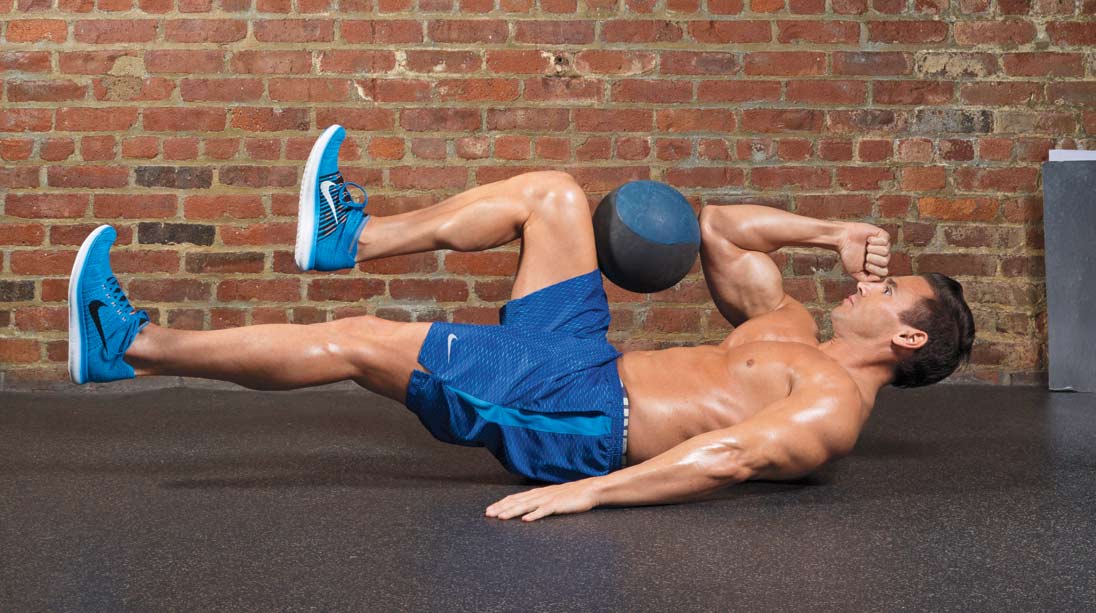
Compresses abdominal contents.
Relations
On the lateral abdominal wall, next to the internal and external abdominal oblique muscles, is the trasversus abdominis. It is located deepest layer of the the lateral abdominal wall.
The transversus abdominis muscle’s aponeurosis also contributes to the rectus sheath’s composition. The largest part of the rectus abdominis and pyramidalis muscles on both their anterior and posterior sides are enclosed by this multilayered aponeurosis.
Together with the aponeurosis of the internal abdominal oblique muscle, the transversus abdominis aponeurosis forms the rear wall of the rectus sheath, which envelops the posterior upper three-quarters of the rectus abdominis. Conversely, the lower fourth of the anterior wall of the rectus sheath is formed by the convergence of the aponeuroses of the transversus abdominis and internal oblique muscle with the aponeurosis of the external abdominal oblique anterior to the rectus abdominis. The arcuate line, which is 2.5 cm below the umbilicus, is the place where they converge.
The transversalis fascia connects to the inguinal ligament and the internal and external oblique below the free bottom border of the transversus abdominis. The transversalis fascia features a circular aperture known as the deep inguinal ring, which is located between the midinguinal point—the halfway point between the ASIS and pubic symphasis—and the midpoint of the inguinal ligament, about 1/1.5 cm above the ligament. The interfoveolar ligament is a band of fibers that occasionally emerges from the free lower border of the transversus abdominis along the medial border of the deep inguinal ring.

8 Comments