Vastus Lateralis Muscle
Table of Contents
Vastus Lateralis Muscle Anatomy
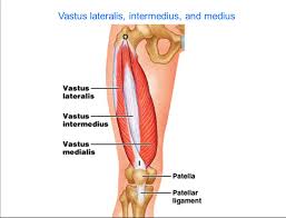
The vastus lateralis muscle is located on the lateral side of the thigh. This muscle is the largest of the quadriceps muscle, which includes: rectus femoris, vastus intermedius, and vastus medialis. Together, the quadriceps act on the knee and hip to promote movement as well as strength and stability.
Origin
It originates from the,
- upper aspect of the intertrochanteric line,
- base of the greater trochanter and onto its anterior surface,
- from the proximal portion of the lateral lip of the linea aspera,
- lateral intermuscular septum.
Insertion
It inserts into the lateral side of the quadriceps tendon, joining with rectus femoris, vastus medialis, and vastus intermedius muscles, enveloping the patella, then by the patellar ligament into the tibial tuberosity.
Nerve supply
The Posterior division of the femoral nerve (L3, L4) supplies the muscle.
Nerve Roots
The affected nerve roots include L2, L3, and L4. L3 is the main nerve root that causes VL activity.
Blood supply
- The superior medial artery is a branch of the lateral circumflex femoral artery.
- The inferior medial artery is a branch of the artery of the quadriceps.
- The lateral artery is actually the first perforator of the deep femoral artery.
Venous Drainage
The perforating veins of the lateral femoral circumflex vein, the deep femoral vein, and additional unidentified veins from the superficial venous circulation are responsible for the venous drainage of the ventricle. The larger named veins in the vicinity that aid in draining have names similar to the matching artery.
Action
It acts as an extensor of the knee.
Functional Contribution of the muscle
In everyday life, the quadriceps muscle group as a whole allows a person to stand up from sitting, and walk up or down stairs, along with basic walking and running. These muscles are not active while standing with knees fully extended, but become active during the heel-strike and toe-off phases of gait.
Vastus Lateralis Muscle Exercises
Strengthening Exercises:
Vastus lateralis muscle exercises are:
1. Step-up Exercises
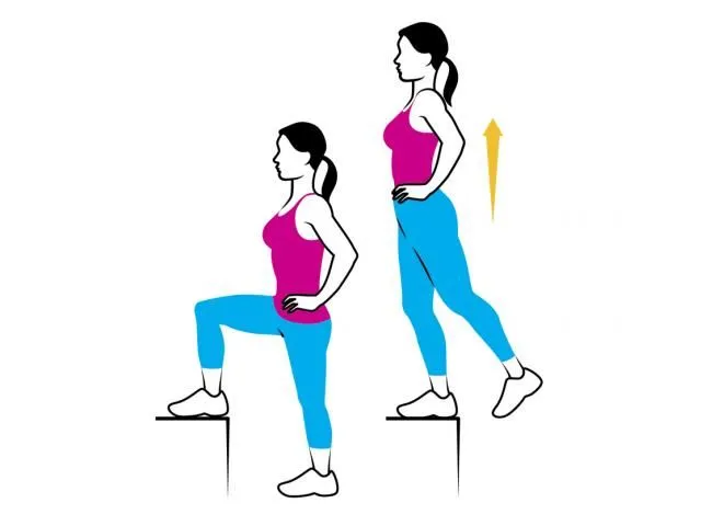
- Start with a box height that is comfortable for you to step up on.
- Be sure to keep your knee in alignment with your second toe.
- Step up and keep your pelvis level and your knee in alignment.
- Be sure to engage the buttocks muscles and fully lock out the knee.
- Return slowly back down to the ground.
- The focus should be on the slow eccentric (lowering) back to the ground for 1 second up and 3 seconds down.
- Perform 2 sets of 15-20 repetitions once per day.
Stretching Exercises:
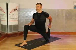
- From a forward lunge position, flex the forward knee with the sole of the foot flat on the floor.
- Flex the knee of the rearward leg towards the buttocks, grasp the foot with your hand behind you and apply a medial rotation to the hip by pushing the foot away from the midline of the body.
- To gently increase the stretch, tilt the pelvis posteriorly and keeping the chest upright, lean into the forward hip.
- You may want to perform this stretch next to a wall or chair to aid in balance.
Anatomical Variations
Nearly 60% of individuals may have two insertional heads on the VL. The vastus lateralis obliquus (VLO) and vastus lateralis long head (VLL) are the names given to these two heads. In most instances, this separation from the longitudinal head is accomplished by a layer of fat or fascia.
However differences between the sites of insertion and origin were rare. The patella’s angulation of fiber insertion varies noticeably from specimen to specimen if the VLO is present. Usually, the VLL inserts at an angle of 10 degrees to 17 degrees, plus or minus 8 degrees. However, the insertional variation of the VLO ranges from 26 to 41 degrees.
Clinical Significance
Some evidence has shown that in patients with patellofemoral pain syndrome vastus lateralis contracts prematurely when compared with vastus medialis which has been hypothesised to be a cause of knee pain. In more recent studies this theory has not been clinically proven and vastus lateralis/medialis timing has been shown to be uneven in healthy test subjects.
Gait Cycle and Vastus Lateralis
The VL, which is a member of the quadriceps muscle group, contracts to prepare the knee for weight bearing toward the end of the swing phase of gait. The muscle group as a whole absorbs most of the force produced by the heel impact. As part of the loading response, the muscle group keeps contracting during the first part of the stance phase. Last but not least, when walking downhill and taking descending steps, the VL, a muscle group belonging to the quadriceps, contracts eccentrically.
Anterior Knee Pain
Because the VL is the strongest muscle in the quadriceps, it plays a major role in anterior knee pain syndromes. About 40% of the total strength of the quadriceps muscle group is thought to come from the VL, 35% from the RF and VI, and the remaining 25% from the VM.
Patellofemoral dysfunction has been linked to both a more proximal attachment of the vastus medialis obliquus (VMO) and overdevelopment of the vastus lateralis. Pain, instability in the joint, and irregular patellar movement can all be signs of a VL-VM imbalance.
Q-angle
The patellar movement could show further abnormalities based on the limb’s Q-angle. The lateral pull of the patella by the VL will have an increased effect on a lower extremity with genu valgum (increased Q-angle), leading to an abnormal stress pattern and acceleration of arthritic processes.
FAQs
On the lateral, or outer, portion of your thigh, is a muscle called the vastus lateralis. The largest muscle in the quadriceps group, it is one of the four muscles in that group. In order to help expand your knee joint, the vastus laterails collaborate with the other quadriceps muscles.
Sudden tension on the muscle, particularly during sports involving lengthening contractions like skiing, frequently activates trigger points in the vastus lateralis muscle. In addition, the muscle is open to direct impact and injury due to its great size and vulnerable position.
It may take up to six weeks or longer to fully recover. Most people recover from a moderate strain or sprain after resting for a week or two. When you are pain-free and have complete range of motion in your leg, your quadriceps will have healed. A gradual rehabilitation program is helpful throughout this period.
Rest, ice, elevation, compression bandaging, ultrasound, massage, and strengthening and stretching activities are some of the treatment options for vastus lateralis pain.
References
Biondi, N. L., & Varacallo, M. (2023, August 8). Anatomy, Bony Pelvis and Lower Limb: Vastus Lateralis Muscle. StatPearls – NCBI Bookshelf. https://www.ncbi.nlm.nih.gov/books/NBK532309/

