
Ankle Joint: Anatomy, Function, Importance
Ankle joints are the most common joints we use in our day-to-day lives, and they’re also the most common joints that require care and attention.
Ankle joints are a series of joints that connect your lower leg to your foot, and they’re used for a wide variety of movements and positions. Even the smallest ankle fractures are painful and difficult to walk on, so it’s important to take care of your ankle joints to prevent future problems. To help keep your ankle joints healthy and strong, try these exercises regularly: squats, calf raises, and lunges.
Table of Contents
Introduction
~This is a synovial joint of hinge variety.
~the bony architecture of the ankle joint:
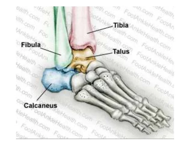
tibia,fibula,talus and calcaneum
Articular surfaces
~Upper articular surface
~inferior articular surface
~superior articular surface
Upper articular surface:
~the upper articular surface is formed by:
~the lower end of the tibia including the medial malleolus.
~the lateral malleouls of the fibula,
~the inferior transverse tibiofibular ligament.
inferior articular surface:
~the stability of the joint is ensured by,
~close interlocking of the articular surfaces.
~strong collateral ligaments on the sides.
~the tendon that crosses the joint, four in front, and five behind.
Superior articular surface:
~the downward projection of medial and lateral malleoli, on the corresponding sides of the talus.
~by the inferior transverse tibiofibular ligament that bridges the gap between the tibia and fibula behind the talus, the socket is provided flexibility by strong tibiofibular ligaments and by slight movements of the fibula at the superior tibiofibular joint.
Ligaments of Ankle Joint:
~the joint is supported by:
~fibrous capsule
~the deltoid or medial ligament
~A lateral ligament
Fibrous capsule:
~it surrounds the joint and is attached all around the articular margins with two exceptions.
(A)Posteriosuperiorly, it is attached to the inferior transverse tibiofibular ligament.
(B)Anteroinferiorly, it is attached to the dorsum of the neck of the talus some distance from the trochlear surface.
~the anterior and posterior parts of the capsule are loose and thin to allow hinge movements, on each side, however, it is supported by strong collateral ligaments.
~the synovial membrane lines the capsule. the joint cavity ascends for some distance between the tibia and the fibula.
Medial ligament:
~this is a very strong triangular ligament present on the medial side of the ankle. the ligament is divided into a superficial and a deep part. both parts have a common attachment, which is indicated by the name of the fibers.
Superficial part:
- Anterior fibers or tibionavicular are attached to the tuberosity of the navicular bone and to the medial margin of the spring ligament.
- The middle fibers or tibiocalcanean are attached to the whole length of the sustentaculum tali.
- The posterior fibers or posterior tibiotalar are attached to the medial tubercle and to the adjoining part of the medial surface of the talus.
Deep part or anterior tibiotalar:
~it is attached to the anterior part of the medial surface of the talus.
~the deltoid ligaments are crossed by the tendons of the tibialis posterior and flexor digitorum longus.
Lateral ligament:
~this ligament consists of 3 bands as follows.
- The anterior talofibular ligament is a flat band that passes from the anterior margin of the lateral malleous to the neck of the talus, just in front of the fibular facet.
2. The posterior talofibular ligament passes from the lower part of the malleolar fossa of the fibula to the lateral tubercle of the talus.
3. The calcaneofibular ligament is a long rounded cord that passes from the notch on the lower border of the lateral malleolus to the tubercle on the lateral surface of the calcaneum. it is crossed by the tendons of the peroneus longus and brevis.
Bone Of Ankle Joint
The true ankle joint is composed of 3 bones.
~tibia and fibula form the superior part.
~tibia which forms the medial portion.
~fibula which forms the lateral portion.
~talus forming the inferior part.
An ankle joint is formed where the leg joins the foot.
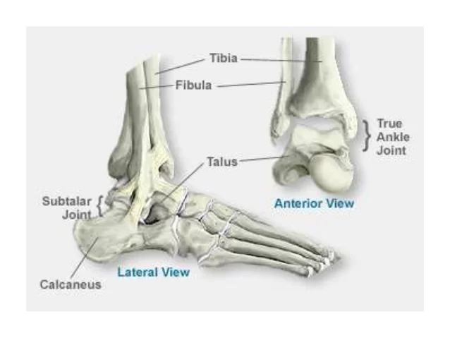
~talo-crural joint.
~crus is leg(knee to foot).
~crura is plural of crus.
~crual is pertaining to the leg.
~it is a synovial hinge joint that connects the distal end of the tibia and fibula with the superior surface of the talus bone.
~the articulation between the tibia and talus bears more weight than between the fibula and talus.
~the articular surface of the tibia is referred to as the plafond.
~plafond(french for ceiling).
~the true ankle joint responsible for
~up(dorsi flexion)
~down(plantar flexion)motion of the foot.
~dorsiflexion consists of the approximation of the dorsum of the foot to the front of the leg.
~angle decreases
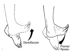
~while in plantar flexion the heel is drawn up and the toes pointed downward.
~the angle increases.
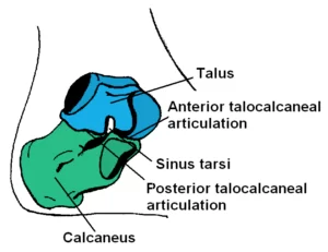
~beneath the true ankle joint is the second part of the ankle, the subtalar joint, which consists of the talus on top and the calcaneus on the bottom.
~the subtalar joint allows side-to-side motion of the foot.
Relations of the knee joint:
~Anteriorly, from the medial to lateral side, there are the tibialis anterior, the the extensor hallucis longus, the anterior tibial vessels, the deep peroneal nerve, the extensor digitorum longus, and the peroneus tertius.
~Posteriorly, from medial to lateral side, there are the tibialis posterior, the flexor digitorum longus, the posterior tibial vessels, the tibial nerve, the flexor hallucis longus, the peroneus brevis, and the peroneus longus.
Blood supply:
~from anterior tibial, posterior tibial, and peroneal arteries.
Nerve supply:
~from deep peroneal and tibial nerves.
Movements of Ankle Joint:
~active movements are dorsiflexion and plantar flexion.
~in dorsi flexion the forefoot is raised, and the angle between the front of the leg and the dorsum of the foot is diminished.
~in plantar flexion, the forefoot is depressed and the angle between the leg and the foot is increased.
Muscles producing movements:
A.Dorsiflexion
Primary muscle: Tibialis anterior
Accessory muscle:
1.extensor digitorum longus
2.Extensor hallucis longus
3.Peroneus tertius
Primary muscle:-1.Gastrocnemius
2. soleus
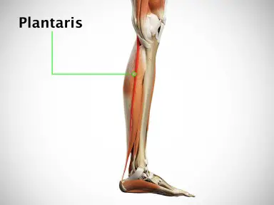
B.plantar flexion
Acessory muscle:
- Plantaris
2. Tibialis posterior
3.Flexor hallucis longus
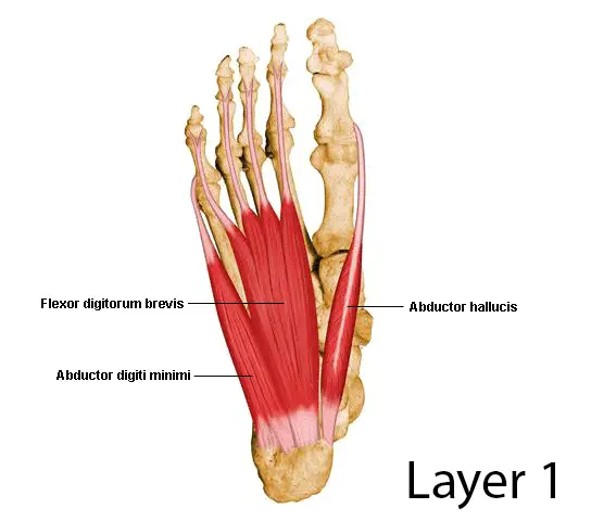
4.Flexor digitorum longus
Clinical Importance of Ankle Joint:
- the so-called Sprains of the ankle are almost always abduction sprains of the subtalar joints, although a few fibers of the deltoid ligament are also torn. true sprains of the ankle joint are caused by forced plantar flexion, which leads to the tearing of the anterior fibers of the capsule.
2. Dislocation of the ankle is rare because the joint is very stable due to the presence of the deep tibiofibular socket. whenever dislocation occurs, it is accompanied by a fracture of one of the malleoli.
3. The optimal position of the ankle (to avoid ankylosis)is one of slight plantar flexion.
~during walking, the triceps surae (plantar flexors) raise the heel from the ground When the limb is moved forward the dorsiflexors help the foot in clearing the ground. the value of the ankle joint resides in this hinge action, in this to-and-fro movement of the joint during walking.
36 Comments