
Flexor Carpi Ulnaris Muscle
Table of Contents
Flexor Carpi Ulnaris Muscle Anatomy
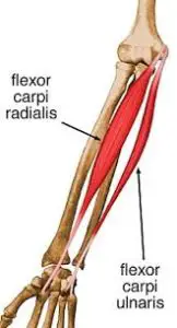
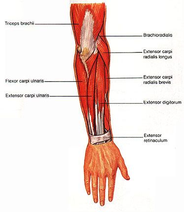
Flexor carpi ulnaris is a fusiform muscle located in the anterior compartment of the forearm. It belongs to the superficial flexors of the forearm. It is the most medial of the superficial flexors.
It is the strongest wrist flexor available. The tendinous arch that connects the two distinct heads of the flexor carpi ulnaris is where it all begins. The ulnar head arises from the olecranon and upper three-fourths of the subcutaneous border of the ulna by aponeurosis, whereas the humeral head arises from a flexor tendon origin on the medial epicondyle.
The fifth metacarpal bone, the hamate hook, and the wrist’s pisiform bone are where the flexor carpi ulnaris inserts. The pisometacarpal ligament allows the flexor carpi ulnaris to insert into the fifth metacarpal bone and the pisohamate ligament into the hook of the hamate.
Origin
It originates from the medial epicondyle of the humerus the medial aspect of the olecranon process and the posterior border of the ulna.
Insertion
It inserts into,
- pisiform bone
- hook of the hamate
- base of the fifth metacarpal bone.
Nerve supply
The ulnar nerve supplies the muscle.
Blood supply
Flexor carpi ulnaris receives its arterial blood supply via three different routes. Proximally, a branch of the posterior ulnar recurrent artery supplies the muscle as it passes between the humeral and ulnar heads. Branches of the ulnar artery supply the middle and distal parts of the muscle, with an accessory supply also present distally via the inferior ulnar collateral artery.
Lymphatic Drainage
The upper limb lymphatic system, which is made up of both superficial and deep lymphatic vessels, includes the flexor carpi ulnaris lymphatic drainage. The cubital lymph nodes, which are close to the medial epicondyle of the humerus, are served by the superficial veins that encircle the basilic vein. The axillary lymph nodes are served by vessels that round the cephalic vein. The major deep veins are followed by the deep lymphatic vessels, which ultimately come to an end in the humeral axillary lymph nodes. They also drain lymph from the flexor carpi ulnaris.
Action
It flexes and adducts the hand at the wrist joint.
Function
Flexor carpi ulnaris can move the hand sideways as well as flex it. Contracting with flexor carpi radialis and palmaris longus, flexor carpi ulnaris produces flexion of the hand at the wrist joint. However, when it contracts alongside the extensor carpi ulnaris muscle in the posterior compartment, its counteracting forces produce adduction of the hand at the wrist, otherwise known as ulnar deviation or ulnar flexion.
Embryology
Around the fourth week, dorsolateral somite cells begin to migrate into the limb to form muscles, giving rise to the upper limb musculature. The muscle tissue separates into distinct extensor and flexor components as the limb buds grow and lengthen, according to connective tissue generated from the lateral plate mesoderm. Fibroblast growth factor ensures Sonic hedgehog expression in cells inside the zone of polarizing activity, so coordinating the anterior-posterior and proximal-distal axis developmental schema.
As a result, the sonic hedgehog makes sure that the apical ectodermal ridge contains the fibroblast growth factor. The ulnar nerve originates from the ventral branch and eventually supplies the flexor carpi ulnaris after the limb buds mature, with the major ventral rami passing through into the mesenchyme.
Strengthening Exercise
1. Towel Crush
- Your forearm should start by resting on the table. You can be seated or standing.
- The towel should be in your hand, rolled up or formed together to make a ball.
- When you‘re ready, squeeze the towel and hold for ten seconds (or as long as you can up to 10 seconds).
- Release and repeat, 3 sets of 10 reps.
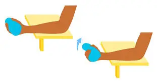
2. Wrist Curves
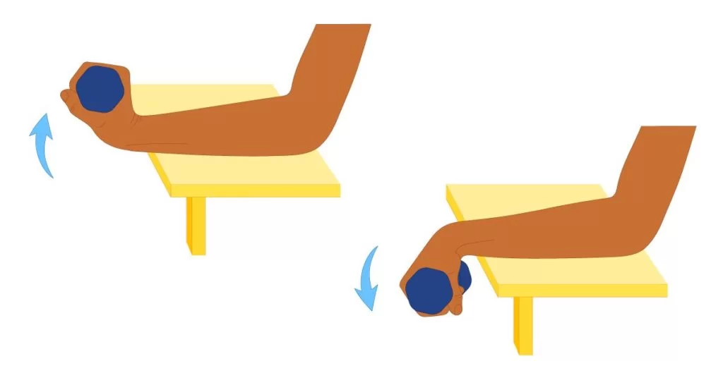
- Sit on a chair or stand near a desktop with a dumbbell in your right hand. Your feet should be about shoulder-width apart.
- Your right arm should be rested on the tabletop with your palm and dumbbell facing up.
- When you‘re ready, lower the dumbbell down to the ground while holding on tightly.
- Once you‘ve reached the lowest point, curl back up to the top of the movement again, contacting the muscles in your forearms. Keep your forearm steady since your wrist is the only part in your arm that should be moving.
- Do a set of ten repetitions and then switch arms. Complete for three sets.
Anatomical Variations
A physiologic variation of the flexor carpi ulnaris, it extends to the radial aspect of the flexor carpi ulnaris and is situated in an anteromedial plane to the flexor digitorium superficialis and between the two. This variation forms a distinct tendon 5 cm above the proximal boundary of the flexor retinaculum, and in one case report, it starts at the medial epicondyle, 1 cm posterolateral to the flexor carpi ulnaris origin. This variation attaches to the wrist bones, including the hamate and triquetral, as well as the flexor retinaculum.
The auxiliary flexor carpi ulnaris has been characterized as having many origins and insertions. The pisiform, the hook of the hamate, the abductor digiti minimi, or the fifth metacarpal can all be inserted by the brevis variation of the accessory flexor carpi ulnaris. It has been discovered that further variations of the auxiliary flexor carpi ulnaris mentioned in the literature insert on the triquetral bone, the flexor pollicus longus tendon, and the flexor digitorum profundus tendon for the index finger.
The ulnar nerve may be compressed within the Guyon canal by malformed anatomical structures, which makes these physiological variations significant. Prior research has revealed a connection between aberrant flexor carpi ulnaris variation in the Guyon canal and ulnar artery thrombosis. In order for healthcare professionals to be ready for altered surgical exposures, it is crucial to be aware of these anatomic variations during preoperative surgical planning.
Stretching Exercise
1. Wrist Flexor Stretch
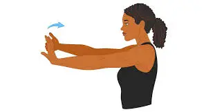
- To stretch flexor carpi ulnaris, begin by holding your right arm straight out in front of you. The palms of your hands should face outwards.
- Take your left hand, hold the fingers of your right hand, and bend them backward. You should feel the stretch on the inside of your right forearm.
- Hold the stretch for 10-15 seconds, repeating as needed.
Surgical Considerations
For several surgical exposures, including the exposure of the ulna’s shaft, the flexor carpi ulnaris is crucial. The main goal of surgery is to expose the ulnar shaft through the inter-nervous plane between the extensor carpi ulnaris and flexor carpi ulnaris muscles when treating for open reduction and internal fixation of ulnar fractures.
The surgeon must be cautious to separate the flexor carpi ulnaris from the ulna epiperiosteally because the ulnar nerve passes beneath it as it goes down the forearm. Before flexor carpi ulnaris is stripped from the ulnar shaft, the surgeon must locate the ulnar nerve between the two heads of the flexor carpi ulnaris. Failure to do so could result in harm to the ulnar nerve.
In order to perform a volar approach to the ulnar nerve at the wrist, the flexor carpi ulnaris is crucial surgically. When the ulnar nerve is compressed, the Guyon canal is decompressed using this volar technique. The volar carpal ligament is thought to be the flexor carpi ulnaris and deep palmar fascia continuing into one another. To expose the ulnar nerve and artery, muscle retraction requires cutting the flexor carpi ulnaris fascia. The ulnar nerve and artery are situated directly below the volar carpal ligament and flexor carpi ulnaris. The ulnar nerve must be protected during both superficial and deep surgical dissection.
The two flexor carpi ulnaris heads are covered by both a deep and superficial aponeurosis, which are freed during subcutaneous transposition of the ulnar nerve for cubital tunnel syndrome. To prevent iatrogenic compression, a transverse fascia arc is located distal to the flexor carpi ulnaris ulnar hiatus and released. When releasing the flexor carpi ulnaris, the surgeon must take care to prevent ligating the motor branches of the flexor carpi ulnaris.
Clinical Importance
Activities such as cutting hair and using tools like a screwdriver or hammer can irritate your muscles and cause flexor carpi ulnaris tendonitis. Sports like swimming, tennis, rock climbing, and rugby can all leave an athlete more prone to developing flexor carpi ulnaris tendonitis due to their use of the FCU.
The ulnar nerve is affected by cubital tunnel syndrome, the second most common compression neuropathy of the upper extremities. The ulnar nerve is most frequently compressed and trapped between the two heads of the flexor carpi ulnaris aponeurosis. There is a space between the two heads of the flexor carpi ulnaris called the cubital tunnel. They are joined by the Osborne ligament, which is the continuation of the fibroaponeurotic covering of the epicondylar groove.
Flexor carpi Paresthesias of the tiny finger, ulnar half of the ring finger, and ulnar dorsal hand may be the result of ulnaris compression of the ulnar nerve; at night, these symptoms may worsen if the arm is propped up. Ulnar nerve compression results from the cubital tunnel flattening during arm flexion when the ligament expands.
Patients may complain of a weak grasp and thumb pinch (Froment’s sign) due to a loss of metacarpal phalangeal joint flexion power and thumb adduction as a result of prolonged compression of the ulnar nerve. Claws on the ring and little fingers, together with muscle atrophy, are signs of chronic compressive syndrome.
Wrist flexion and ulnar deviation can duplicate the focal volar and ulnar-sided wrist discomfort that is the hallmark of flexor carpi ulnaris tendinopathy, an overuse injury. A sesamoid found in the Flexor carpi ulnaris tendon, the pisiform is one of the factors that causes wrist tendinitis.
3 Comments