
Pronator Teres Muscle
Table of Contents
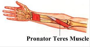
Pronator teres Muscle Anatomy
The pronator teres muscle is located on the palmar side of the forearm, below the elbow. It is a fusiform muscle found in the anterior forearm.
It can be found on the front of the forearm and is a long, spherical muscle. The humeral head and the ulnar head are the two origins of this muscle.
The Humeral head is a more superficial and large in size as compared to Ulnar head which is deeper and joints the both head at an acute angle, making a single muscle.
Sometimes the ulnar head is missing, other times it is more muscular or tendinous.
In 83% of arms, the median nerve travels between the two pronator teres heads, where it is frequently compressed. The medial border of the Cubital Fossa is formed by the pronator teres.
Origin
It originates from the medial epicondyle of the humerus.
- Origin of Humeral Head: the deep antebrachial fascia, the common flexor tendon, and the medial epicondyle of the humerus.
- Origin of Ulnar Head: medial aspect of the ulna’s coronoid process.
Insertion
It inserts into the middle of the lateral aspect of the shaft of the radius.
Nerve supply
The median nerve supplies the muscle.
Blood supply
- Branches of ulnar artery; common interosseus artery, anterior ulnar recurrent artery
- Branch of the radial artery; radial recurrent artery
- Branches of brachial artery; inferior ulnar collateral arteries.
Action
The pronator teres pronates the forearm and helps the elbow joint bend. Together with the pronator quadratus, it has a combined effect. The muscles are shortened and less able to generate force when the elbow is completely extended.
Antagonist Muscle:
Supinator, Biceps Brachii Muscle
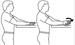
Strengthening exercise
Grasp a dumbbell with your thumb against the inner surface. Sit on a bench or chair, bend your arm, and place your upper arm on the backrest with your thumb facing upward. Rotate the dumbbell so your thumb is facing down, then return to your starting position. Repeat for your desired number of repetitions then switch arms.
Stretching Exercises:
The action of the muscle is to pronate the forearm.
1. Pronator Stretch:
- Bend one elbow next to your body and place the other hand on the back of your hand.
- With help from the other hand, rotate your forearm to bring the palm of your hand facing the ceiling until you feel a stretch in the forearm.
- Hold the stretch for 20-30 seconds.
- Maintain the position and relax.
Related pathology
Pronator teres syndrome is a compression neuropathy of the median nerve at the elbow. It is rare compared to compression at the wrist (carpal tunnel syndrome) or isolated injury of the anterior interosseous branch of the median nerve (anterior interosseous syndrome).
The most common cause is entrapment of the median nerve between the two heads of the pronator teres muscle. Other causes are compression of the nerve from the fibrous arch of the flexor superficialis, or the thickening of the bicipital aponeurosis.
FAQ
As per name suggests, the pronation of the forearm—an exclusive upper limb movement—is the major function of the pronator teres. At the proximal radioulnar joint, the muscle rotates the proximal section of the ulna by pulling the radius medially.
the median nerve
The median nerve (MN), which travels between these heads, innervates pronator teres muscle. The “PT syndrome” (nerve entrapment), which might happen in this phase, is possible. Potential risk factors for this condition could be anatomical differences in this area.
originates from the coronoid process of the ulna and the medial epicondyle of the humerus. It attaches laterally to the radius’s middle shaft.
3 Comments