Risorius Muscle
Table of Contents
Risiorius Muscle Anatomy
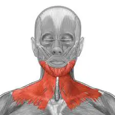
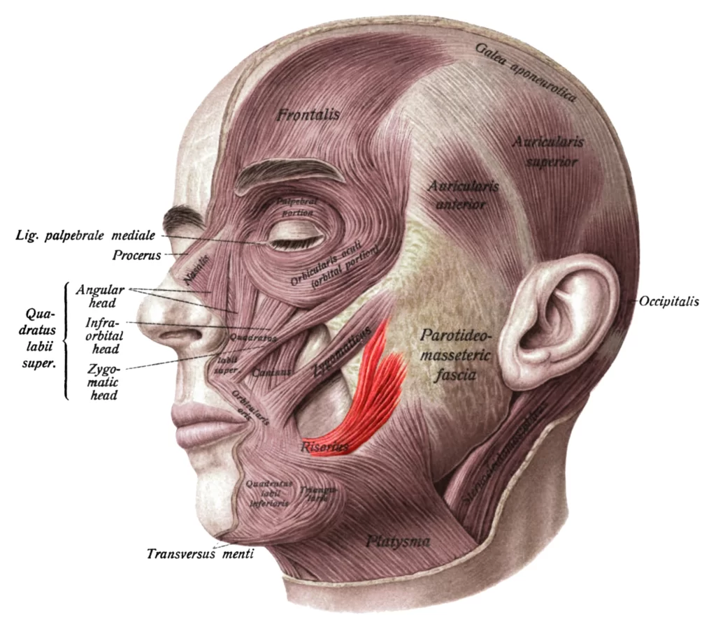
The Risorius begins around the parotid gland, a salivary gland in the back of the jaw, and wraps around the platysma muscle, a muscle located in the chest and neck. Smiling and frowning are two of the facial expressions that are created by all of the facial muscles working together.
Origin
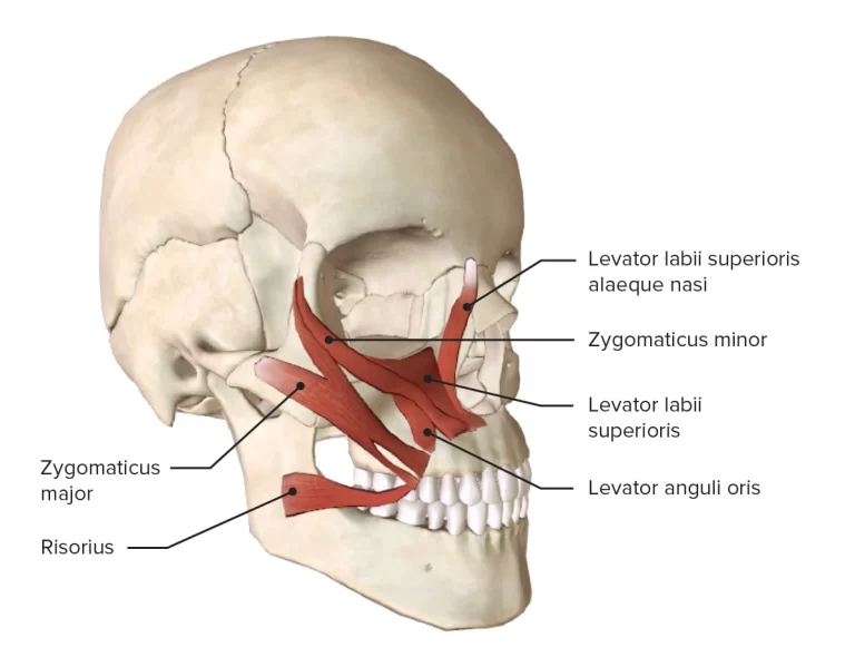
It originates from the masseteric fascia.
The risorius muscle fibres originate at these sites and converge medially, travelling in a nearly horizontal direction towards the mouth’s angle. The modiolus is a thick, movable, fibromuscular mass that is formed when the risorius interlaces with other muscles that converge towards the mouth’s angle.
Insertion
It inserts into the skin of the angle of the mouth.
Nerve supply
The buccal branch of the facial nerve supplies the muscle.
Blood Supply
The facial artery and the transverse facial artery supply the arterial blood to the muscle.
Risorius Muscle Action
Upon activation, the risorius pulls the angle of the mouth laterally. The risorius retracts the angle of the mouth to produce a smile, albeit an insincere-looking one that does not involve the skin around the eyes.
Compare this with a real smile, which raises the lips with the action of zygomaticus major and zygomaticus minor muscles and causes “crow’s feet” around the eyes using the orbicularis oculi muscles.
Relations
Risorius runs along the extension of the line connecting the mandibular and maxillary alveolar processes in the superficial layer of the facial muscles. Overlying the masseter muscle and the buccopharyngeal fascia that divides it from the buccinator muscle, it is enclosed in the superficial fascia of the cheek. The facial artery, which gives rise to the superior labial artery, one of its main branches, passes between the risorius and buccinator muscles.
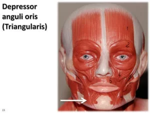
The parotid duct punctures the buccinator muscle to enter the oral cavity, and the parotid gland is connected to the proximal part of the risorius. It blends with the zygomaticus major superiorly, the depressor anguli oris inferiorly, and the orbicularis oris laterally as it gets closer to the angle of the mouth, aiding in the creation of the modiolus.
Embryology
Approximately during the fourth week of gestation, the second pharyngeal arch mesoderm gives birth to the risorius muscle and its artery supply.
Variation
The risorius muscle varies a lot. It can take the shape of a wide, narrow fan. It could be asymmetrical and missing in a sizable proportion of individuals.
Exercises for Risorius muscle
1. Smile Exercise
- Stretch the corners of your mouth laterally (to the sides) while keeping your lips together; hold for 10 seconds.
- Expand the lateral stretch and part your lips to expose the edge of your teeth; hold for 10 seconds.
- Stretch further laterally and expose about half of your teeth; hold for ten seconds.
- Smile as wide and hard as you can, with all your teeth showing; hold for ten seconds.
- Repeat steps 3, 2, and 1 to reverse the smile gradually.
Related pathology
In Bell’s palsy risorius muscle commonly paralysis.
Other facial expressive muscles, including the risorius, may be impacted in Bell palsy, a disorder caused by a deficiency in the facial nerve that manifests as paresis or paralysis of the muscles it innervates.
While the combination of antivirals, such valacyclovir, plus steroids is a debated therapy option, antivirals by themselves have not demonstrated any advantage over a placebo. Physical Therapy treatment may be required. Bell palsy can mimic the symptoms of tumours, Lyme disease, and stroke, thus clearing these other conditions out during the workup may be necessary.
FAQ
To activate the risorius muscle you can perform the exercise – Place an index finger on both corners of the mouth while the lips are fully closed. Contracting the cheek muscles, try to give a big smile without separating the lips.
Risorius is a voluntary muscle of the facial group of muscles that is also part of the buccolabial group of muscles.