
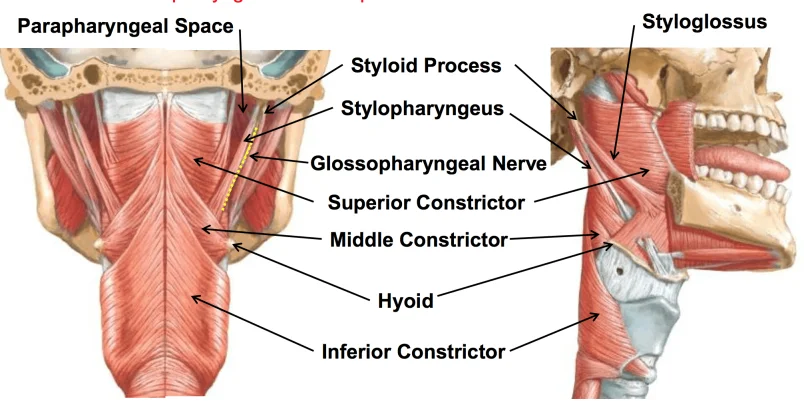
Stylopharyngeus Muscle
Table of Contents
Stylopharyngeus Muscle Anatomy
The stylopharyngeus muscle is a long, slender muscle located in the pharynx, which is the muscular tube connecting the nasal and oral cavities to the larynx and esophagus.
The stylopharyngeus is a muscle in the head that stretches between the temporal styloid process and the pharynx.
The stylopharyngeus is a long, slender muscle, cylindrical above, and flattened below.
It arises from the medial side of the base of the temporal styloid process, passes downward along the side of the pharynx between the superior pharyngeal constrictor and the middle pharyngeal constrictor, and spreads out beneath the mucous membrane.
Some of its fibers are lost in the constrictor muscles while others, joining the palatopharyngeus muscle, are inserted into the posterior border of the thyroid cartilage.
The glossopharyngeal nerve runs on the lateral side of this muscle and crosses over it to reach the tongue.
Origin
- Styloid process (temporal).
Insertion
- thyroid cartilage (larynx).
Nerve Supply
Actions
- elevate the larynx, elevate the pharynx, and swallow.
Blood Supply
- Pharyngeal branch of ascending pharyngeal artery.
Lymphatic drainage
The middle cervical lymph nodes that drain into the supraclavicular lymph nodes moderate the lymphatic outflow of the stylopharyngeus muscle area.
Function
- elevates the larynx
- elevates the pharynx
- dilates the pharynx to permit the passage of a large food bolus, thereby facilitating swallowing.
- Resistance is used depending on the condition of the patients.
Structure
A long, lean, and tapered pharyngeal muscle is the stylopharyngeus. It is flattened inferiorly and cylindrical superiorly.
Before spreading out beneath the mucosal membrane, it travels inferior-ward down the pharynx’s side between the middle and superior pharyngeal constrictors, which are both located superficially to the stylopharyngeus.
Relations
The most medial and vertical of the three styloid muscles is the stylopharyngeus.
The muscle is located in the middle of the internal and external carotid arteries.
It is located anterior to the buccopharyngeal fascia and posterior to the superior constrictor muscle on the lateral pharyngeal wall.
This muscle’s lateral side is where the glossopharyngeal nerve travels before crossing over to the tongue.
Variation
There could be extraneous muscles present that come from neighboring parts of the cranium and could have clinical significance.
Development
The third pharyngeal arch is the embryological origin. It begins to develop between the fourth and seventh weeks of pregnancy.
FAQs
A long, lean, and tapered pharyngeal muscle is the stylopharyngeus. It is flattened inferiorly and cylindrical superiorly.
Before spreading out beneath the mucosal membrane, it travels inferior-ward down the pharynx’s side between the middle and superior pharyngeal constrictors, which are both located superficially to the stylopharyngeus.
The stylopharyngeus muscle is not located in the larynx; rather, it is a muscle of the pharynx, specifically the pharyngeal constrictors.
The function of the stylopharyngeus muscle is to elevate and widen the pharynx during swallowing. It plays an important role in the process of deglutition (swallowing) by assisting in the movement of the bolus (chewed food) through the pharynx and into the esophagus. Additionally, the stylopharyngeus muscle is involved in the process of speech, as it helps elevate the larynx and widen the pharynx, contributing to the modulation of sound during speech production.
A muscle in the pharynx is called the stylopharyngeus. It differs from other pharyngeal muscles in that the glossopharyngeal nerve, not the vagus nerve, innervates it.
Glossopharyngeal nerve (CN IX) innervation.
Due to the embryological development and anatomical placement of the nerves in the head and neck region, the glossopharyngeal nerve (cranial nerve IX), which supplies the stylopharyngeus muscle, is a part of the head and neck.
Early in embryonic development, particular pharyngeal arches give rise to distinct head and neck structures. The third pharyngeal arch, also referred to as the glossopharyngeal arch, is where the stylopharyngeus muscle originates. The third pharyngeal arch is also the primary source of cranial nerve IX, the glossopharyngeal nerve.
The nerve that nourishes the components that grow from a certain pharyngeal arch also originates from that arch. The “arch-to-nerve” rule is a phenomena that causes this. Since the stylopharyngeus muscle comes from the third pharyngeal arch, the glossopharyngeal nerve, which transports motor fibers to control the muscle’s contraction, innervates the muscle.
4 Comments