External intercostal muscles
External intercostal muscles are eleven in number on both sides.
origin:
Lower border of ribs.
Insertion :
Upper border of rib below.
Nerve:
intercostal nerves.
Actions:
Inhalation.
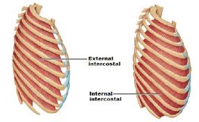
External intercostal muscles are eleven in number on both sides.
origin:
Lower border of ribs.
Insertion :
Upper border of rib below.
Nerve:
intercostal nerves.
Actions:
Inhalation.
Physiotherapist , Samarpan Physiotherapy Clinic, Vastral, Nirant Cross Road, Ahmedabad
Home Visit Treatment Also Available in Bapunagar Vastral Rabari Colony Char Rasta, CTM, Maninagar , Viratnagar , Nikol Nava Naroda And NearBy Area Of Ahmedabad.
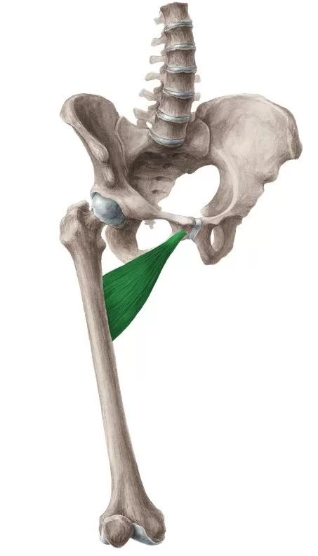
Adductor brevis Muscle Anatomy Adductor brevis is a flat, triangular muscle that is found in the inner thigh. Together with the adductor longus, adductor magnus, gracilis, and pectineus muscles, it comprises a group of muscles known as the adductors of the thigh. Origin It originates from the inferior ramus and body of the pubis. Insertion…
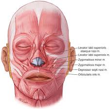
Levator Labii Superioris Muscle Anatomy Origin The levator labii superioris muscle has three points of origin that blend and passes into the upper lip. All three origins attach together within the upper lip and at the time of contraction, raise the upper lip. A strap of the angular head attaches within the ala of the…
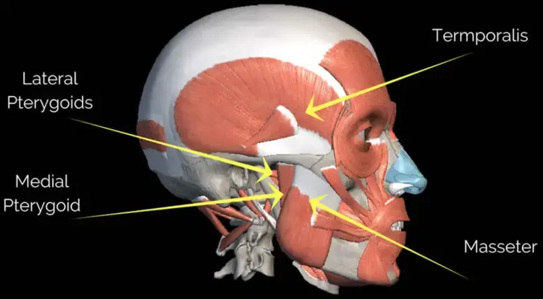
Introduction The masseter is a quadrilateral muscle that covers the lateral surface of the ramus of the mandible. The masseter muscle fibres are arranged in three layers. The masseter muscle is a paired, strong, thick, and rectangular muscle that originates from the zygomatic arch and extends down to the mandibular angle. It contains a superficial…
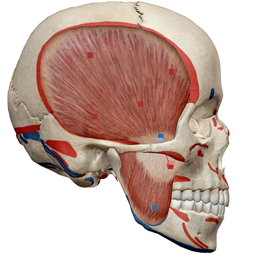
Introduction The muscles of mastication move your mandible during mastication and speech. The muscles of mastication are the Masseter, the Temporalis, The Lateral pterygoid, and the medial pterygoid. They develop from the mesoderm of the first branchial arch and are supplied by the mandibular nerve which is the nerve of the branchial arch. The muscles…
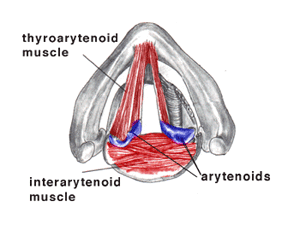
Thyroarytenoid muscle is a broad, thin muscle that forms the body of the vocal fold and that supports the wall of the ventricle and its appendix. It functions to relax the vocal folds. It is part of the intrinsic musculature of the larynx and plays a crucial role in vocal production and control. origin :…
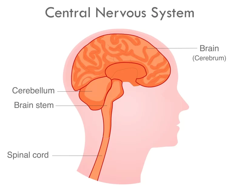
What is a Central Nervous System (CNS)? The central nervous system (CNS) is a complex network of the brain and spinal cord that coordinates and regulates the functioning of the entire body. It plays a critical role in processing sensory information, controlling movements, and executing cognitive functions. The CNS consists of two main components: the…