semitendinosus muscle
| | |

Semitendinosus Muscle Anatomy, Function, Exercise

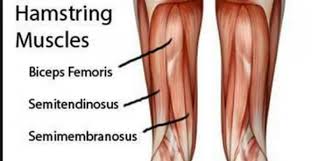
Semitendinosus Muscle Anatomy Semitendinosus muscle is one of 3 Hamstring group muscles that are located backside of the thigh and is responsible for the flexion of the knee. Other Hamstring muscles are the biceps femoris and semimembranosus. All three muscles (semimembranosus, semitendinosus, and biceps femoris) are part of the hamstring muscles. Semitendinosus and Semimembranosus are…

Middle Pharyngeal Muscle
| |

Middle Pharyngeal Muscle

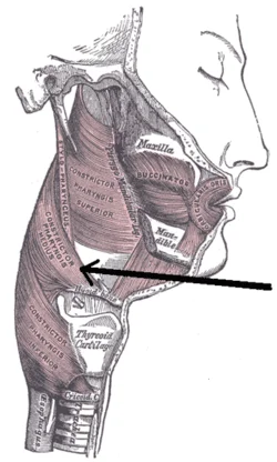
Middle Pharyngeal Muscle Anatomy The middle pharyngeal constrictor is a fan-shaped sheet-like muscle of the pharynx. This muscle, along with the superior and inferior pharyngeal constrictors, forms the bulk of the pharyngeal wall. The pharyngeal constrictor muscles create a peristaltic wave that facilitates passage of the food bolus inferiorly, from the pharynx towards the esophagus,…

Zygomaticus major muscle
| |

Zygomaticus Major Muscle

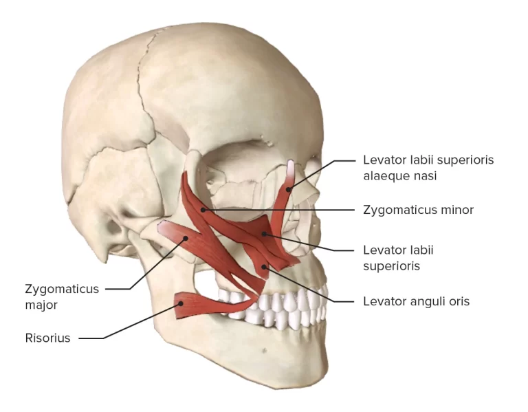
Anatomy of Zygomaticus Major Zygomaticus major shown in red. It is a muscle of facial expression which draws the angle of the mouth superiorly and posteriorly to allow one to smile. Origin: Anterior surface of zygomatic bone. Insertion: Modiolus at angle of mouth. Nerve supply: Buccal branch of facial nerve (VII). Blood supply: Superior labial…

The Cerebrum

Introduction: ~the cerebrum is made of two cerebral hemispheres which are incompletely separated from each other by the median longitudinal fissure.~the two hemispheres are connected to each other across the median plane by the corpus callosum. each hemisphere contains a cavity, called the lateral ventricle. External feautures: three surfaces: 1. the superolateral surface is convex…

The Cerebellum: Anatomy, Function

Introduction: ~The cerebellum is the largest part of the hind brain. it is situated in the posterior cranial fossa behind the pons and medulla.~it is an infratentorial structure that coordinates voluntary movements of the body. Relations: ~Anteriorly:-fourth ventricle,pons and medulla~Posteroinferiorly:-Squamous occipital bone,&~Superiorly:-tentorium cerebelli External features: ~the cerebellum consists of two cerebellar hemisphere that is united…

spinal cord

Spinal Cord

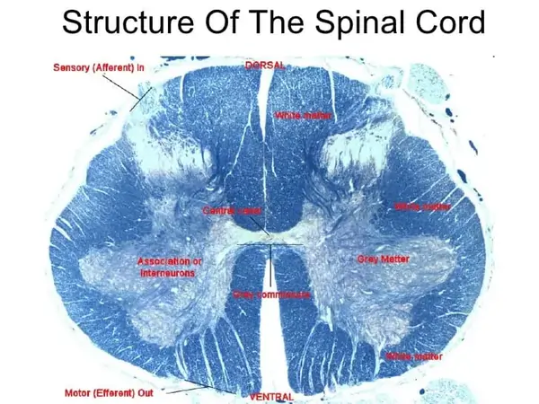
What is the spinal cord? ~the spinal cord is the lower elongated, cylindrical part of the CNS. It occupies the upper two-thirds of the vertebral canal. It extends from the level of the upper border of the atlas to the lower border of vertebra L1 or the upper border of vertebra L2. ~it is about…

Iliococcygeus muscle
| |

Iliococcygeus Muscle

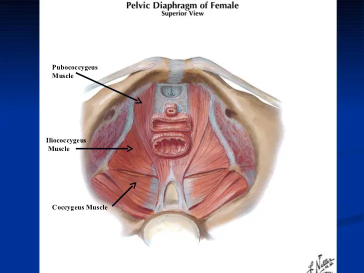
Iliococcygeus Muscle Anatomy The levator ani muscle, which makes up the majority of the pelvic diaphragm, is composed of three muscles, including the iliococcygeus muscle. The flat, broad skeletal muscle is known as the iliococcygeus. the inner side of the ischium (the lower and back part of the hip bone) and from the posterior part…

|

Ankle Joint: Anatomy, Function, Importance

Ankle joints are the most common joints we use in our day-to-day lives, and they’re also the most common joints that require care and attention. Ankle joints are a series of joints that connect your lower leg to your foot, and they’re used for a wide variety of movements and positions. Even the smallest ankle…

Internal oblique muscle
| |

Internal oblique muscle: Origin, Insertion, Function, Exercise

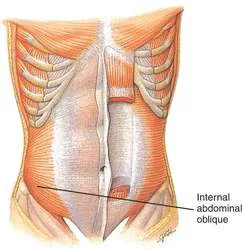
Internal oblique muscle is a thin, broad muscle in the lateral abdominal wall that is located below the external oblique and just above the transverse abdominal muscles. Starting from the superficial layer to the deep layer, the external oblique, internal oblique and transversus abdominis make up the three layers of the lateral abdominal wall. Internal…