

Semitendinosus Muscle Anatomy, Function, Exercise
Table of Contents
Semitendinosus Muscle Anatomy
Semitendinosus muscle is one of 3 Hamstring group muscles that are located backside of the thigh and is responsible for the flexion of the knee.
Other Hamstring muscles are the biceps femoris and semimembranosus. All three muscles (semimembranosus, semitendinosus, and biceps femoris) are part of the hamstring muscles.
Semitendinosus and Semimembranosus are located on the medial side of the posterior thigh. It is a long muscle that reaches the full length of the Back of the thigh – from the hip to the knee.
Origin of the Semitendinosus muscle:
The muscle originates with the long head of the biceps femoris, from the lower medial facet of the lateral section of the ischial tuberosity.
It is also originated from the medial surface of the tendon of the long head of the biceps femoris and also originates from the ischial tuberosity with a thin tendon and a muscular part.
Insertion:
The muscle inserts at the upper part of the medial surface of the tibia, behind the attachment of sartorius and infero-anterior to the attachment of gracilis.
Nerve Supply:
The tibial portion of the sciatic nerve (L5, S1, 2).
The semitendinosus muscle is innervated by the tibial nerve, which originates from the sciatic nerve. Sacral plexus branches into the sciatic nerve, which includes nerve roots extending from L4 to S2, in close proximity.
After passing through the posterior compartment, the sciatic nerve splits into the tibial and common fibular nerves. The semitendinosus and every other muscle in the posterior compartment of the thigh, with the exception of the short head of the biceps femoris, are innervated by the tibial nerve, either before to or following its split from the common fibular nerve.
Blood supply:
The internal iliac, popliteal, and profunda femoris arteries Branches.
The profunda femoris artery, or deep artery of the thigh, is the greatest branch of the femoral artery and provides the majority of the circulatory supply to the semitendinosus muscle. The circulatory supply of the muscle is specifically provided by the profunda femoris artery’s perforating branches.
Venous Drainage
Similar to this, the profunda femoris vein’s perforating veins, which empty into the femoral vein, are responsible for the venous drainage of the semitendinosus muscle.
Function of the Semitendinosus muscle:
The main function is the flexion of the knee with other Hamstring muscles such as biceps femoris, and semimembranosus
Extension of the Hip with gluteus maximus, semimembranosus, biceps femoris (long head), and adductor magnus (posterior part).
It is also a weak internal rotator of the Hip.
Internal rotation of the knee when the knee is flexed with other Agonists muscles such as popliteus and semimembranosus while Antagonist muscles are biceps femoris (long head) and biceps femoris (short head) Sartorius and gracilis assist with the medial rotation of the knee.
Anatomical Variation
Anatomical variations of the semitendinosus muscle have been identified in case studies. One such instance occurred during reconstructive surgery for the anterior cruciate ligament, when the semitendinosus muscle released an extra tendinous attachment during the graft harvesting process.
The muscle released an additional tendinous slip at the insertion location of the semitendinosus muscle, which was connected to the gracilis tendon. With a second tendinous implantation into the semitendinosus tendon, the gracilis did the same. Consequently, a “double” pes anserinus was present. Additionally, investigations have indicated that the semitendinosus tendon inserts into the leg’s crural fascia instead of the superior anteromedial tibia.
Muscle Relations
In the posterior compartment of the thigh, the semitendinosus muscle travels alongside the semimembranosus and biceps femoris muscles. The hamstring muscle complex is made up of these three muscles. The most medial muscle of the posterior compartment of the thigh is the semimembranosus muscle, which is located deep in the semitendinosus muscle.
The posterior oblique ligament, the arcuate ligament, the posterior joint capsule, and the medial tibial condyle are where the semimembranosus muscle inserts after emerging from the superolateral portion of the ischial tuberosity.
The muscle known as the biceps femoris is the most lateral muscle in the posterior compartment. Its small head rises from the lateral lip of the femur’s linea aspera, while its long head originates from the ischial tuberosity. After that, the biceps femoris inserts into the lateral condyle of the tibia and the head of the fibula.
Embryology
The ectoderm, endoderm, and mesoderm are the three germ layers that emerge during the embryo’s gastrulation phase. With the exception of the skull, the paraxial mesoderm specifically develops into striated skeletal muscle and the skeleton.
Thus, the mesoderm is the source of both the muscle and the bone insertions of the semitendinosus muscle. By the conclusion of the fourth week of the embryonic phase, the lower limb as a whole starts to form. As the embryo enters the fetal phase in the eighth week, the limb continues to develop and becomes well-differentiated.
Clinical importance:
A grafted semitendinosus tendon to repair cruciate or collateral ligament injury is mostly used in arthroscopic knee surgery. This choice provides fewer post-operative complications to kneeling.
Semitendinosus strain is a mostly overused sports injury, most common in sports like football, Hockey, and rugby where running, and jumping with suddenly jerky knee movements are associated. The most common treatment is the use of the RICE Principle with the use of Ice packs and Hot packs. After a week gradual strengthening and stretching exercises of the muscles are the best options to return to sports.
One of the most common muscle conditions among athletes is hamstring strains, which are associated with extended periods of time away from competition. Most injuries happen when you accelerate quickly or run at a high speed. Although hamstring strains have also been linked to the semitendinosus muscle, the biceps femoris is typically the muscle in the posterior compartment that sustains injuries the most frequently.
Exercises for semitendinosus:
There are mainly 2 types of exercise – strengthening exercise and stretching exercise.
How to stretch semitendinosus?
Following are the best exercises you can do at home to stretch the muscles.
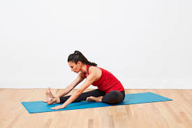
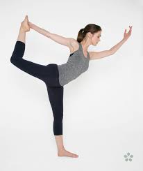
Semitendinosus stretch:

You can take a long sitting position on the soft mat with both legs out straight.
Try to reach at the toes by extending the arms as seen in the pictures- as far as possible by bending at the waist while keeping the knees straight.
Hold this position for 10 to 15 seconds.
Relax back into the first position.
Repeat 2-3 times.
you will feel a gentle stretch at the back of your thighs. Avoid painful exercise.
Hurdler Hamstring stretch exercise:
This is the Best exercise for semitendinosus muscle stretch.
- You can take a long sitting position on the soft mat with both legs out straight.
- Bend one leg at the knee and position the sole of that foot against the opposite inner thigh.
- Try to reach at the toes by extending the arms as seen in the pictures- as far as possible by bending at the waist while keeping the knees straight.
- Hold this position for 10-15 seconds.
- Then return to the first position.
- Repeat with the other leg.
Strengthening exercise of the semitendinosus
To strengthen your semitendinosus muscles, you can do this exercise.
Hamstring curls with Gym Ball exercise:
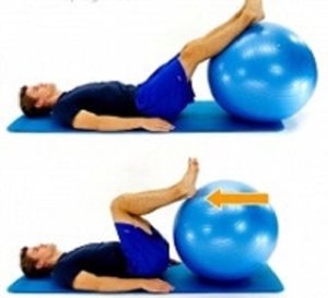
Take a supine lying position on the soft mat with the feet and lower legs resting on a gym ball with knees straight and arms placed over the side of the body.
Gradually curl the ball towards the body by flexing the knees and then gradually roll the ball away.
Repeat this exercise 10-15 times.
Static Hamstring contraction:
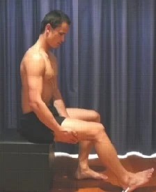
To do this exercise, you have to take a sitting position with the knee bent to about 45 degrees.
Press the heel into the floor by contracting the back of your Hamstring.
Hold for 5-10 seconds and repeat 10 times (exercise must be pain-free)
Surgical Importance
For ACL restoration, the semitendinosus and gracilis muscle tendons are frequently utilized as an alternative to the bone-patellar-tendon-bone graft (BPTB). The sartorius fascia is cut during grafting for ACL restoration in order to reveal the gracilis and semitendinosus tendon underneath. Because these two tendons are not attached to the bone outside of their attachment sites, it is possible to distinguish them from the medial collateral ligament, which is located deep within the gracilis and semitendinosus muscles.
The initial diameter of the gracilis and semitendinosus tendons determines the hamstring graft’s diameter, unlike the BPTB graft. The surgeon can measure the graft’s diameter in a BPTB graft as the graft is being harvested. The BPTB graft contains bone, in contrast to the hamstring autograft, which is thought to enhance graft integration.
Initially thought to be the gold standard for ACL restoration, the BPTB graft is the most often used autograft in the United States. Which autograft is better, though, is up for discussion. Reduced osteoarthritis, donor site morbidity, and anterior knee pain are advantages of hamstring autografts. Benefits of the BPTB autograft include a higher percentage of patients recovering to preinjury activity levels, quicker graft integration, and a lower chance of rupture. Both graft options have modest failure rates, while BTBP has a lower failure rate. As a result, using the gracilis and semitendinosus tendons as an autograft is still a viable alternative for reconstruction, however it should be considered individually.
According to MRI studies, 75% of patients have their semitendinosus tendon regrow after harvesting, and the remaining 25% of patients see a compensatory increase in the size of their semimembranosus muscle. In addition to their frequent application in ACL restoration, semitendinosus and gracilis tendon grafts have also been used to treat large rotator cuff tears that have previously seen muscular atrophy, degeneration, and retraction.


8 Comments