Cricothyroid Muscle
The cricothyroid muscle is the only tensor muscle of the larynx aiding with phonation.
The cricothyroid muscle is a little muscle located deep in the anterior compartment of the neck that is bilaterally paired. It is one of the six intrinsic laryngeal muscles, along with the other six:
- Posterior and lateral cricoarytenoids
- oblique and transverse arytenoids
- thyroarytenoid
- aryepiglotticus
- thyroepiglotticus
- vocalis muscles.
The cricothyroid muscle’s primary job is to aid in vocalization.
Origin:
Anterior and lateral cricoid cartilage .
Insertion:
Inferior cornu and lamina of the thyroid cartilage.
Nerve Supply:
External branch of the Superior laryngeal nerve (branch of the vagus nerve).
The cricothyroid muscle receives motor innervation from the vagus nerve (CN X) via the external branch of the laryngeal nerve. As far as the intrinsic muscles of the larynx are concerned, only this one is not supplied by the recurrent laryngeal nerve.
Blood Supply
The cricothyroid artery provides the cricothyroid muscle with its arterial supply. The superior thyroid artery, a branch of the external carotid artery, gives origin to this artery. The anterior cricothyroid ligament is where the cricothyroid artery crosses superiorly to meet the contralateral cricothyroid artery.
Lymphatic Drainage
The cricothyroid muscle is drained by a vein of the same name. It is a branch of the internal jugular vein that empties into the superior thyroid vein.
Actions:
tension and elongation of the vocal folds.
Function
Rotation about the cricothyroid joint results from the contraction of the cricothyroid muscle, which pulls the thyroid cartilage anteriorly and downward. As a result, the cricoid and thyroid cartilage are closer together, and the thyroid cartilage is moved away from the arytenoid cartilage. The result is a thinning, tightness, and stretching of the vocal folds. Higher-pitched sounds are produced during vocalization by longer, tighter vocal folds.
Structure
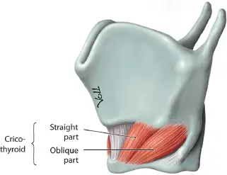
This little muscle is located between the thyroid and cricoid cartilages of the larynx, deep in the anterior neck. It starts in the anterolateral region of the cricoid cartilage arch. After that, the muscle fibers split into two groups: oblique and straight.
The oblique portion that is placed inferiorly moves posterolaterally before inserting into the thyroid cartilage’s inferior horn, or cornu. The straight portion inserts onto the inferior margin of the thyroid cartilage lamina by traveling posterosuperiorly at an angle higher than the oblique part’s fibers.
Muscle Relations
There are a number of significant neighbors for every cricothyroid muscle that are helpful clues for surgical planning. First, the cricothyroid ligament occupies a triangle that divides it from its contralateral counterpart.
When constructing an emergency airway, this is a crucial location. The muscle lies medial to the cricothyroid joint and caudal to the inferior margin of the thyroid cartilage. It passes deep to the inferior constrictor muscle and its related tendinous arch, but superior to the lateral cricoarytenoid muscles. Adjacent to the cricothyroid muscle are the superficial infrahyoid muscles.
As the muscle moves toward the midline, the cricothyroid artery and the superior laryngeal nerve’s external branch approach it at a superolateral angle. The cricothyroid muscles are also partially covered by the superior poles of the ipsilateral lobe and, if present, the pyramidal lobe of the thyroid gland.
Clinical Importance
A cricothyroidotomy may result in damage to the cricothyroid muscle. When alternative methods cannot be used to produce a patent airway, this emergency surgical treatment is used. A surgical incision is created between the superior margin of the cricoid cartilage and the base of the thyroid cartilage, through the cricothyroid ligament, anteriorly. This airway is intended to be used as a temporary until a more permanent one can be created.