
Duchenne muscular dystrophy (DMD): Cause, Symptoms, Physiotherapy Treatment
Duchenne muscular dystrophy (DMD), an incurable inherited neuromuscular disorder is the most common of the more than 30 types of muscular dystrophy. It is a genetic disease that leads to progressive deterioration of muscle fibers of the body.
The condition usually affects boys only which is one of the most frequent genetic conditions affecting approximately 1 in 3,500 male births worldwide but girls can also carry the mutated gene and feel some symptoms. They have half times the risk of passing the mutated gene on to their sons, who will be affected by the disease.
Table of Contents
Causes
DMD is caused by a mutation in the DMD gene, which encodes for a protein called dystrophin, which is the largest gene identified in humans and is located in the short arm of the X chromosome.
There are various types of mutations associated with DMD, with the majority leading to no means fail, or very little, functional dystrophin being produced.
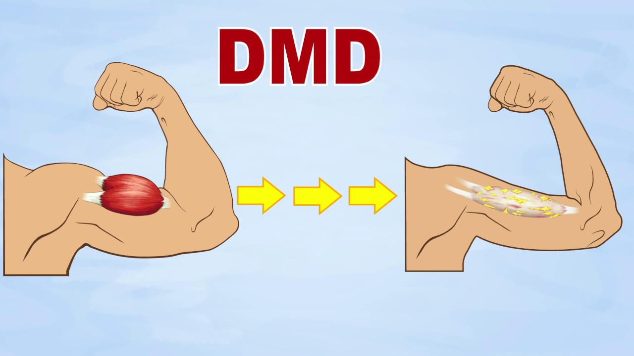
Dystrophin is primarily found in the skeletal muscles, which are responsible for movement, and in the heart or cardiac muscle, a protein that is essential to the proper functioning of our muscles. Without dystrophin, muscles are not able to function or repair themselves properly which means the loss of muscle then results in a loss of strength and function.
Dystrophin acts as an anchor, keeping muscle cells altogether and connecting them with the body of the surrounding part. It normally functions as giving strength and protection of muscles from damage while they are contracting and relaxing work.
The absence of dystrophin sets in motion a cascade of harmful effects on muscles. With the increase of time, damaged cells become weakened and die, and are replaced by scar tissue, leading to muscular weakness. Fibrous tissue commencing to form in the muscle, and the body’s immune system increases inflammation. This protein keeps muscle cells intact. Its absence leads to rapid muscular deterioration as a child with DMD grows.
Signs and Symptoms
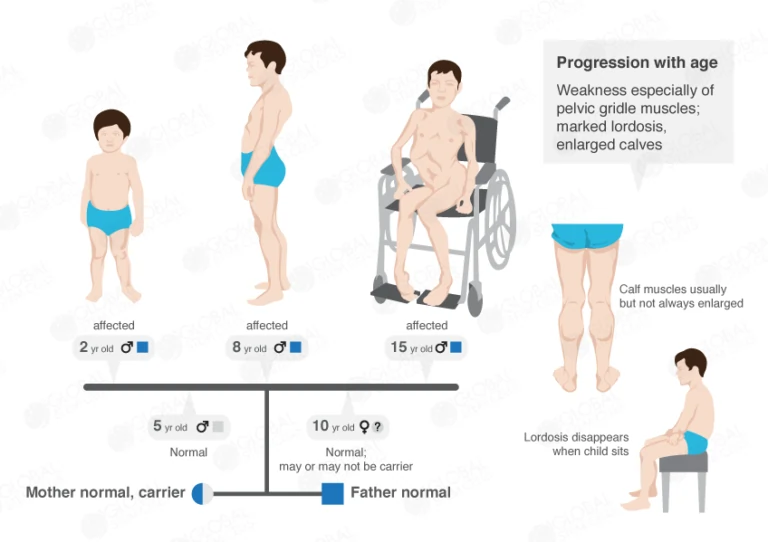
- Duchenne muscular dystrophy is the most commonly seen and severe form of the disease. Although girls can be carriers who are sometimes mildly affected, it’s much more common in boys. It usually starts when a child is between ages 2 and 5 typically appears in early childhood.
Following are the most Symptoms of Duchenne muscular dystrophy include:
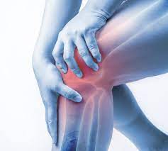
- Muscle weakness that begins in the hips, pelvis, and legs are related to Duchenne muscular dystrophy (DMD) selectively affects the limb muscles close to the trunk before those which ones far from it as well as the legs are affected before the arms.
- Muscle pain and stiffness along with muscle cramps at times.
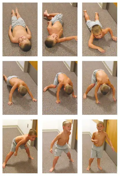
- Difficulty in standing includes while getting off from the floor affected boys may use own hand support to push themselves to an upright position known as Gower’s maneuver
- Trouble learning to sit independently and walk
- Trouble running and jumping
- Unsteady, waddling gait. To try to keep their balance, they may stick out their tummies and pull back their shoulders. Children also have difficulty in raise their arms.
- The flexed joint that cannot be straightened known as flexion contracture
- Clumsiness, falling frequently
- Walking by using the toes or balls of the feet instead of using the heel
- trouble in rising from a lying or sitting position
- difficulty in climbing upstairs
- Larger-than-normal calves are sometimes painful which enlargement is known as pseudohypertrophy, or “false enlargement,” because the muscle tissue is abnormal.
- Trouble breathing
- Learning disabilities or behavioral problems with cognitive disability.DMD occurs in three general areas: emotional interaction, verbal learning, and memory attention focusing
- Deficiency of speech and language development
- The curvature of the spine “scoliosis” due to can cause one hip to rise higher than the other.
- Because of Lack of dystrophin, the muscle layer in the heart (myocardium) can become weak, a condition is known as cardiomyopathy
- Generalized hypotonia means decreased muscle tone
- a child may not have symptoms like complain of shortness of breath, problems that indicate poor respiratory function which includes headaches, mental dullness, difficulty concentrating or staying awake, and nightmares. Breathing problems that may sometimes require the use of a ventilator
- Growth velocity with DMD is generally slower than normal in the early years of life, which leads to short stature.
- By age 12, most children with Duchenne muscular dystrophy must use a wheelchair to get surrounding site.
- The disease also damages the heart muscles along with muscles that are needed to breathe, which can be life-threatening. Patients with DMD often die in their late teenage years or 20s by conditions called respiratory insufficiency or cardiomyopathy; only a few DMD patients survive beyond 30 years.
Diagnosis
A diagnosis of DMD is made based upon a thorough clinical evaluation, a detailed patient and family history, and performing a physical examination, this doctor may find pseudohypertrophy, lumbar spine deviation, gait abnormalities, and several grades of diminished muscle reflexes. Along with that, a variety of specialized tests including molecular genetic tests can be done.
If the genetic tests are not knowledgable, surgical removal and microscopic examination include biopsy of affected muscle tissue that may reveal characteristic changes to muscle fibers. Specialized blood tests such as creatine kinase that evaluate the presence and levels of certain proteins in the muscle known as immunohistochemistry are also used.
Blood tests :
CK and other enzyme levels
It reveals elevated levels of creatine kinase (CK). Early in the diagnostic procedure, doctors frequently order a blood test named a CK level. CK stands for creatine kinase, an enzyme that leaks out and is found in abnormally high levels when the muscle is damaged.
When elevated CK levels are detected in a blood sample, it usually means the muscle is being disintegrated by an abnormal process, for instance, muscular dystrophy or inflammation. A very high CK level suggests about the muscles themselves and not the nerves that control them are the likely cause of the weakness, however, it can not indicate exactly what type of muscle disorder might be happening.
The detection of higher CK levels which is usually in the thousands or ten thousand range can confirm that muscle is damaged or inflamed, but cannot confirm a specific diagnosis of DMD. High levels of CK may be detected before the onset of signs and symptoms, even in infants affected by DMD.
The CK level reaches up to 10 to 20 times the upper limit value by age 2, then progressively declines at a rate of 25% annually, eventually returning to normal level when a considerable amount of muscle tissue has been replaced by fat and scar or fibrotic tissue.
Genetic testing
Generally, genetic diagnosis is suggested for patients with elevated serum CK levels and clinical diagnosis of dystrophinopathy. Diagnosis is confirmed if a mutation of the DMD gene is recognized. The genetic analysis is initially directed to find large deletion or duplication mutations. If the initial genetic analysis is negative, the analysis of small and microdeletion/duplication gene mutations is after then.
Muscle biopsy
To gain more information on the patient, a doctor may order a muscle biopsy, the surgical removal of a small sample of muscle from the patient. By examining this collection of samples, doctors can tell a great affection about what is actually happening inside the body muscles.
Modern technology may use the biopsy to identify muscular dystrophies from inflammatory and other disorders and to differentiate among different forms of muscular dystrophy. For example, the amount of functional dystrophin protein recognized in a muscle biopsy sample light without dystrophin present.
Cardiac investigation
A doctor may sometimes observe characteristic changes associated with conduction abnormalities in an electrocardiogram in patients with Cardiomyopathy with DMD. Also, structural changes in the heart, as valvular heart disease, especially affecting the mitral valve when it occurs can be detected by echocardiography. So that, noninvasive imaging with echocardiography, electrocardiograms, and cardiac MRI are important with the consultation of a cardiologist.
Treatment
There is no known treatment for Duchenne muscular dystrophy (DMD) condition. Treatment is mainly focused on managing the symptoms of DMD and related complications caused by severe progressive muscle weakness and loss. Medications, for example, steroids may improve the strength and function of muscles.
These can help improve muscle power and function. An enlarged, weakened heart which means dilated cardiomyopathy may be treated with medications, but in severe cases, a heart transplant may be recommended. Assistive devices like inhalers for breathing difficulties may be needed, especially at night and as the disease progresses.
Specialists who may be involved in the care of patients with DMD may include :
- Neurologist
- Orthopedist
- Cardiologist
- Pulmonologist
- pediatrician
- medical geneticist
- Medical geneticist
- Physiotherapist
- Occupational therapist
Physiotherapy Treatment in Duchenne muscular dystrophy:
Physiotherapy Treatment will be a significant part treatment plan from the time of diagnosis of the condition. It is vital that you have specialized physical therapy evaluations every 4-6 months. By this, any and all changes can be recognized over time and needs can be addressed instantly.
In the initial stages of the condition, the physiotherapist will be involved in helping keep the child active and normal while during later stages of the condition, the physiotherapist will help more with respiratory issues as well.
Physiotherapists play an important role in:
- Reduce contractures by introducing regular stretching into your daily routine
- Maintain function and adapt to any loss of function
- Monitor function over time through standard tests and measures
- Assess for and manage compromised skin integrity
- Prevent and manage pain
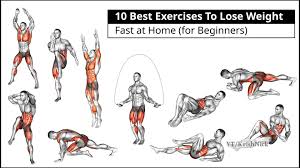
- Prescribe exercise and supervision safe physical activity that is, low load, low resistance exercise
- Recommend mobility devices, adaptive seating, and other equipment
- Rehabilitation after injury or fracture
Physiotherapy is significant to the treatment of Duchenne’s Disease. It is important to monitor regularly the physical signs and symptoms of the condition and physiotherapy can help keep the child active for as long as time possible. Physiotherapists will work with the parents and caregivers and provide them with information and manual skills which will be helpful for the child.
Exercise for DMD
Contractures are one of the major complications, which is a physiotherapist will treat. They will do these by a stretching exercise, which can also be taught to the parents or caregivers. Stretching of all joints including the shoulder, elbow, wrist, ankle, knee, and hip should start as early as possible and continue through adulthood.
Potential advantages of stretching may include:
- Temporary improvement of blood flow to the muscle
- Decrease discomfort
- A feeling of well-being and good
- Temporary increase in tolerance to stretch
- A temporary decrease in muscle stiffness
- Continued movement by the joint’s full available range of motion.
Physiotherapists will also be responsible for suggesting the parents on any orthoses, such as Ankle foot orthosis (AFOs), and referring them to a pediatric orthopedist. They will also help families with mobility aids and equipment the child may need.
Physiotherapists monitor the child’s posture in sitting, lying, and standing position. They teach the parents various ways to help the child sit, stand, and lie in optimal positions using pillows or splints. Asleep system and night splints can be recommended for nighttime to help maintain the child’s posture over a long period of time.
Due to the muscle wasting caused by the lack of dystrophin protein, DMD patients may struggle with many everyday activities. Physiotherapists can help with the treatment of neuromusculoskeletal problems.
They can help slow the regression of range of motion, muscle strength, power and endurance, daily function, work to improve gait pattern and posture or alignment. Physiotherapy can also reduce the pain that the patient may be experiencing.


2 Comments