Geniohyoid muscle
Table of Contents
Introduction
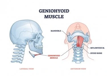
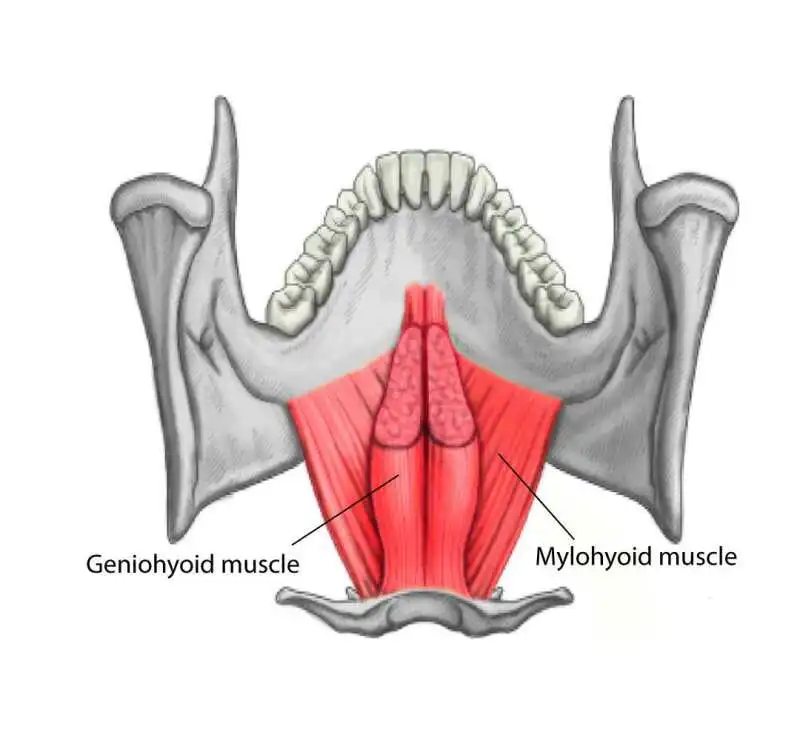
Geniohyoid muscle is a short and narrow muscle that lies above the medial part of the mylohyoid muscles.
The Geniohyoid muscle is a short, paired muscle that belongs to the suprahyoid muscle group of the neck. Together with the digastric, stylohyoid, and mylohyoid muscles, it makes the floor of the oral cavity.
The geniohyoid muscle’s main function is to coordinate the movements of the floor of the mouth and the hyoid bone while swallowing and voice production.
Origin
The geniohyoid muscle originated From the inferior mental spine (genial tubercle) of the mandible.
Insertion
The geniohyoid muscle fibers run backward and downwards to be inserted into the anterior surface of the body of the hyoid bone.
Nerve supply
The first cervical nerve. the muscle fibers pass through the hypoglossal nerve.
Blood supply
The blood supply of the geniohyoid muscle is the Sublingual branch of the lingual artery
Action
- Elevates the hyoid bone.
- It May depress the mandible when the hyoid bone is fixed.
Clinical Relevance of Geniohyoid muscle
- Conditions that can affect the mylohyoid muscle include:
- Overuse injuries
- Strains
- Myopathy
- Atrophy
- Infectious myositis
- Neuromuscular diseases
- Contusions
- Lacerations
Exercise for Geniohyoid muscle
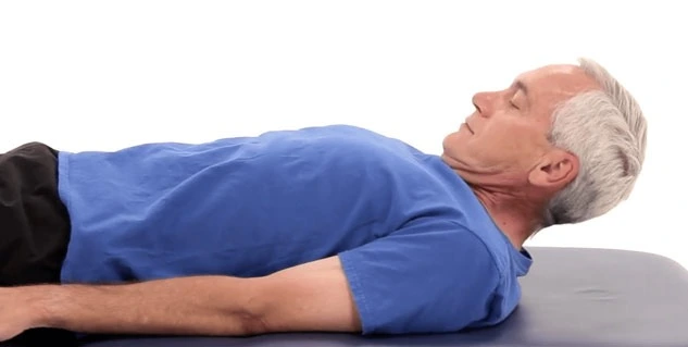
This exercise is used to strengthen the suprahyoid muscles, including the geniohyoid. Strengthening these muscles may relieve dysphagia. This exercise has an isometric and an isokinetic portion.
Lay flat on the ground and don’t place a pillow under your head as you need it to be completely flat. Lift your head while keeping your both shoulders firmly on the ground. Tuck your chin into your chest and hold this position for 30 seconds then rest. Repeat this exercise three times.
FAQs
The short, paired geniohyoid muscle is a member of the neck’s suprahyoid muscle group. It forms the oral cavity’s floor together with the digastric, stylohyoid, and mylohyoid muscles.
One of these muscles is the paired mylohyoid, which also includes the geniohyoid, digastric, and stylohyoid muscles. These muscles affect the hyoid bone’s mobility, which is why they are commonly referred to as the supporting muscles of mastication.
The geniohyoids are two little paired bundles that start at the posterior mentum and insert on the smaller cornu of the hyoid’s anterior side.
A little paired muscle called the geniohyoid is located above the mylohyoid muscle’s medial border. Its journey from the chin to the hyoid bone is whence it gets its name (“genio-” is a common prefix for “chin”).
During swallowing, the geniohyoid muscle contracts, pulling the hyoid bone and the connected larynx upward and forward. The geniohyoid muscles work in concert with the mylohyoid muscles to depress the mandible and draw it inward, opening the mouth, but when the hyoid bone is fixed, the mouth is opened.

