Lower Back Muscle Pulled
Table of Contents
Introduction
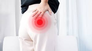
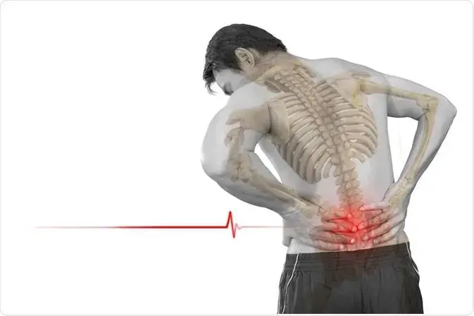
Lower back muscle pulls, also known as strains, can be a source of significant discomfort and limited mobility. The lower back, or lumbar region, is a complex network of muscles, ligaments, and joints that support the spine and facilitate various movements. When these muscles are subjected to excessive force, overuse, or improper lifting techniques, it can lead to strain and injury.
This condition is not uncommon and can affect people of all ages and activity levels. Whether it’s due to sudden movements, poor posture, or repetitive stress, a pulled lower back muscle can result in pain, stiffness, and difficulty performing everyday activities.
In this exploration of lower back muscle pulls, we will delve into the causes, symptoms, and common treatments for this condition. Understanding the factors contributing to a pulled lower back muscle is crucial for prevention and effective management.
Anatomy of the Back Muscle
Your back is made up of numerous muscles. Some muscles support your trunk and spine. Others support your breathing, help you stand up straight, and move your body.
Injuries to the back muscles are frequent because they bear the brunt of your weight support and are involved in a wide range of motions. Low back pain can result from these injuries. You should warm up before physical activity and maintain the strength of your other muscles to prevent injury and maintain the health of your back muscles.
Other Terminology
Since the superficial and intermediate muscle groups—also known as extrinsic muscles—represent upper limb muscles that have moved to the back during foetal development, they are also referred to as immigrant muscles. True back muscles are another name for the deep/intrinsic back muscles. The thoracolumbar fascia divides them from the extrinsic muscles, which are situated deep in them.
Physiotherapy’s Significance
Correct diagnosis and treatment of back pain require a thorough understanding of the anatomy of the back muscles. A common presenting symptom for clients is generalized back pain, which is typically caused by a strain in the skeletal muscle.
There are three categories of back muscles: superficial, intermediate, and deep.
- Superficial
It’s Connected to shoulder motions.
Intermediate: connected to thoracic cage movements.
Deep: connected to spinal column movements.
Intrinsic muscles are those that originate in the back during the embryonic stage. The muscles that are categorized as extrinsic are the superficial and intermediate muscles, which do not originate in the back.
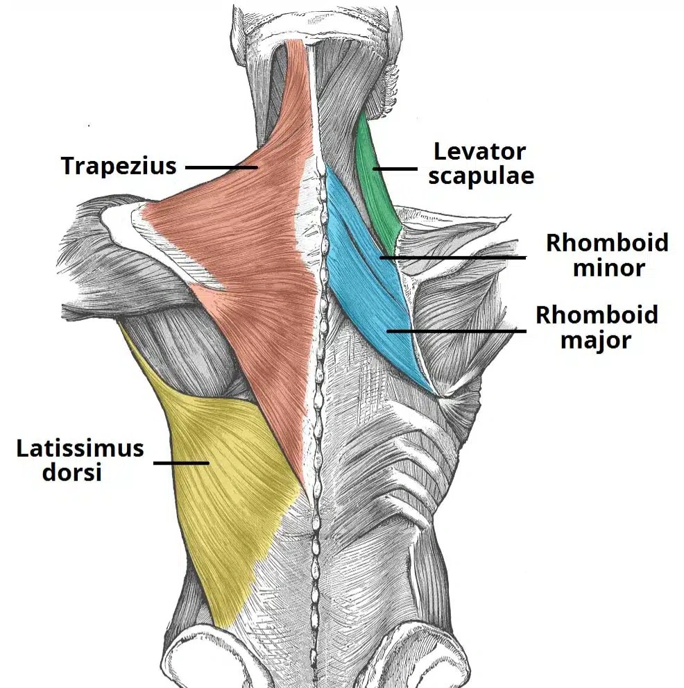
The attachments, innervations, and functions of the superficial back muscles are discussed in this article. Underneath the skin and superficial fascia are the superficial back muscles. They attach to the clavicle, scapula, and humerus, the shoulder bones, after emerging from the vertebral column. As a result, every one of these muscles is connected to motion.
The latissimus dorsi, rhomboids, Levator scapulae, and trapezius are the muscles that make up this group. The most superficial muscles are the latissimus dorsi and the trapezius, with the trapezius covering the Levator scapulae and rhomboids.
- Trapezius
The muscle known as the trapezius is broad, flat, and triangular. A trapezoid shape is formed by the muscles on either side. Out of all the back muscles, it is the most superficial.
Attachments: Derives from the ligamentous nuchal, the spinous processes of C7–T12, and the skull. The fibres are affixed to the scapula spine, acromion, and clavicle.
Innervation: The accessory nerve provides motor innervation. Proprioceptor fibres from the C3 and C4 spinal nerves are also sent to it.
Actions: During arm abduction, the upper fibres of the trapezius raise and rotate the scapula.
- Latissimus Dorsi
The latissimus dorsi has its origin in the lower back, spanning a large region.
Attachments: Widely distributed; originates from the inferior three ribs, iliac crest, thoracolumbar fascia, and spinous processes of T6–T12. The fibres come together to form a tendon that joins to the homerun’s intertubercular sulcus.
The thoracodorsal nerve is innervated.
Actions: The upper limb extends, adducts, and rotates medially.
- Levator Scapulae
The Levator scapulae is a tiny muscle that resembles a strap. It starts in the neck and moves downward to join the scapula.
Attachments: Joins the medial border of the scapula to the transverse processes of the C1–C4 vertebrae.
The dorsal scapular nerve is innervated.
Actions: Raise the shoulder blades.
- Rhomboids
The major and minor rhomboid muscles are two in number. The major is positioned inferior to the rhomboid minor.
Rhomboids major
Attachment: Originates from the spinous processes of the T2-T5 vertebrae are the Rhomboid Major Attachments. connects to the scapula’s medial border, in the space between the inferior angle and the scapula spine.
The dorsal scapular nerve is innervated.
Actions: Scapula rotation and retractions.
Minor Rhomboids
Attachment: Derived from the spinous processes of the C7-T1 vertebrae. attaches at the level of the scapular spine to the medial border of the scapula.
The dorsal scapular nerve is innervated.
Actions: Scapula rotation and retractions.
2. Intermediate Group
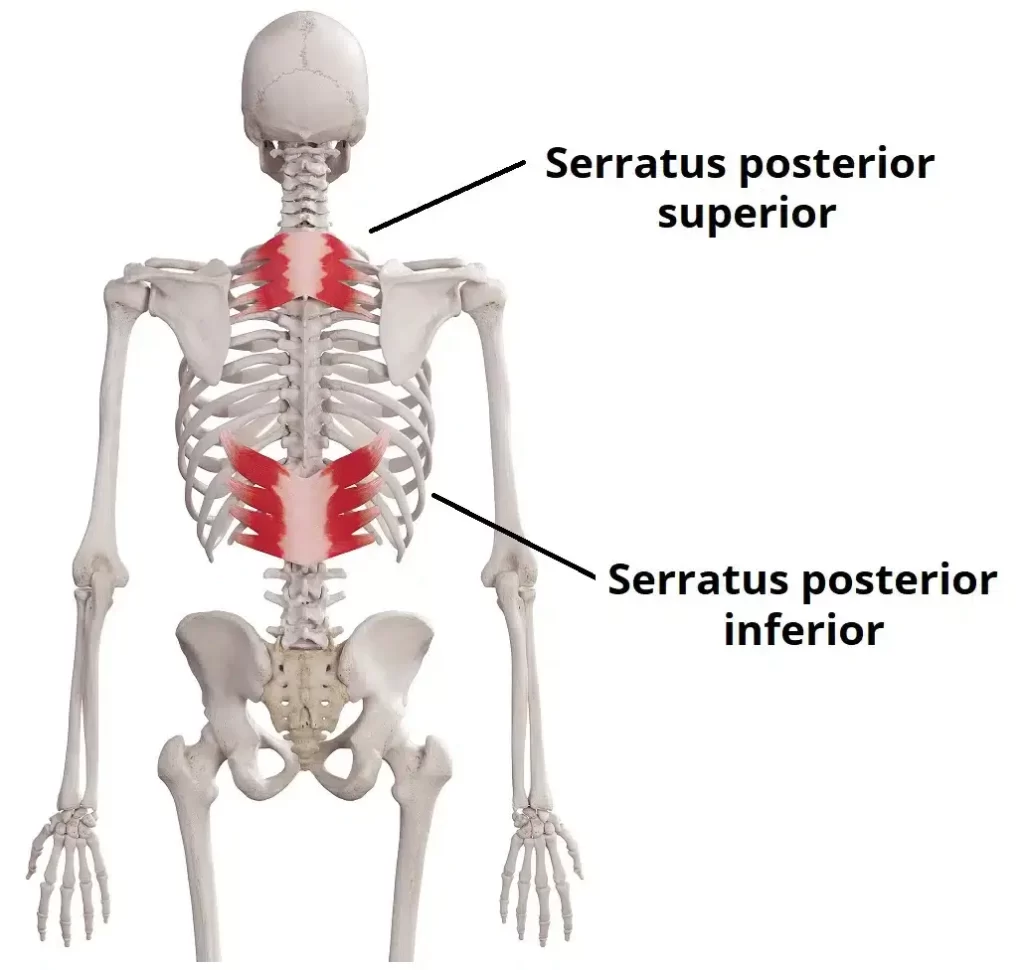
- Serratus Posterior Superior
Attachments: Originates from the lower part of the ligamentous nuchal, and the cervical and thoracic spines (usually C7 – T3). The fibres pass in an inferolateral direction, attaching to ribs 2-5.
Actions: Elevates ribs 2-5.
Innervation: Intercostal nerves.
- Serratus Posterior Inferior
This muscle is broad and strongest. It lies underneath the latissimus dorsi.
Attachments: They arise from the thoracic and lumbar spines (generally T11 – L3). The fibres pass in a super lateral direction, attaching to ribs 9-12.
Actions: Depresses ribs 9-12.
Innervation: Intercostal nerves.
3. Deep – it’s connected to spinal column movements.
Intrinsic muscles are those that originate in the back during the embryonic stage. The muscles that are categorized as extrinsic are the superficial and intermediate muscles, which do not originate in the back. The attachments, innervations, and functions of the deep (intrinsic) back muscles are discussed in this article. Together, the well-developed deep back muscles stretch from the sacrum to the base of the skull. They are connected to the regulation of posture and the motions of the spinal column.
Deep fascia, the layer covering the muscles, is essential to their structure. Deep, intermediate, and superficial back muscles are the three layers that make up the deep back muscles anatomically.
The splenius capitals and splenius services are the two muscles in this group. They are both connected to head and neck motions. They cover the deeper neck muscles and are situated on the posterolateral aspect of the neck.
- Capitals Splenius
Attachments: Derives from the spinous processes of vertebrae C7 through T3/4 and the lower aspect of the ligamentous nuchal. The fibres affix to the occipital bone of the skull as well as the mastoid process.
Innervation: C3 and C4 spinal nerves’ posterior rami.
Action: Move your head to the same side as your action.
- Splenius Cervices
Attachments: Protrudes from the T3–T6 vertebrae’s spinous processes. The fibres connect to C1-3/4’s transverse processes.
Innervation: The lower cervical posterior rami
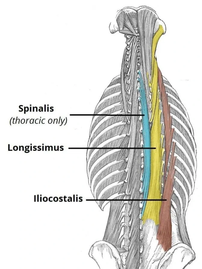
Action: Move your head to the same side as your action. In between Three intrinsic back muscles are located in the intermediate region: the iliocostalis, longissimus, and spinals. The erector spinal is a column made up of these muscles combined. The vertebral spinous processes and the ribs’ costal angle are located posterolateral to the spinal column, where the erector spinae is located. By their superior attachments, all three muscles can be further divided into the lumbar, thoracic, cervices, and capitals. Additionally, they all share a Tendinous origin that originates from:
- Iliocostalis – The erector spinae contains the iliocostalis muscle laterally. It has three parts: lumbar, thoracic, and crevices, and it is related to the ribs.
Origin: from the common tendinous origin and attaches to the cervical transverse processes as well as the costal angle of the ribs.
Innervation: The spinal nerves’ posterior rami.
Actions: Laterally flexes the vertebral column by unilateral action. extends the head and spinal column bilaterally.
- Longissimus – The iliocostalis and spinal muscles are separated by the longissimus muscle. Out of the three columns, it is the biggest. It is separated into three sections: the capitals, cervices, and thoracic.
Attachments: Originating from the common tendinous origin, it is attached to the mastoid process of the skull, the transverse processes of C2 through T12, and the lower ribs.
Innervation: The spinal nerves posterior rami.
Actions: Laterally flexes the vertebral column by unilateral action. extends the vertebral column bilaterally.
- Spinalis – Located medially within the erector spinae is the Spinalis muscle. Out of the three muscle columns, it is the smallest. It is separated into three sections: thoracic, services, and capitals (some people do not have the services portion).
Attachments: Grows from the common tendinous origin and joins the occipital bone of the skull, the spinous processes of C2, and T1–T8.
Innervation: The spinal nerves’ posterior rami.
Actions: Laterally flexes the vertebral column by unilateral action. extends the head and spinal column bilaterally.
The transversospinales are the collective name for the deep intrinsic muscles that are situated beneath the erector spinae. These are a collection of short muscles connected to the spinous and transverse processes of the spinal column. This group of muscles consists of the rotators, multifidus, and semispinalis.
- Semispinalis – Of the deep intrinsic muscles, the semispinalis is the most superficial. It can be separated into thoracic, cervices, and capital muscles based on its superior attachments, just like the intermediate muscles.
Attachments: C4-T10 transverse processes are the source of origin. The fibres attach to the occipital bone of the skull as well as the spinous processes of vertebral segments 4-6.
Innervation: The spinal nerves’ posterior rami.
Actions: The head and spinal column are extended and rotated contra laterally.
- Multifidus – The semispinalis muscle is situated beneath the multifidus. The lumbar region is where it is most advanced.
Attachments: Originates from the sacrum, posterior iliac spine, erector spinal’s common tendinous origin, lumbar vertebrae’s mamillary processes, T1–T3 transverse processes, and C4–C7 articular processes. Its origin is broad. The fibres cling to the spinal spinous processes as they ascend 2-4 vertebral segments.
Innervation: The spinal nerves’ posterior rami.
Actions: Promotes spinal column stability.
- Rotators – The muscles in the transversospinales group that are the deepest are the rotators. The thoracic region is where they are most noticeable.
Attachments: Derived from the transverse processes of the vertebrae. The fibres climb up and affix to the spinous processes and lamina of the higher vertebrae.
Actions: aids in the vertebral column’s rotation and extension. additionally serves to stabilise the vertebrae and has proprioceptive effects.
Innervation: The spinal nerves’ posterior rami
- Small Deep Internal Muscles
Spans between neighbouring spinous processes are known as interspinal. helps to keep the spinal column stable. Spans between neighbouring transverse processes are known as intertranversarii. helps to keep the spinal column stable.
- Lavatories costarum
Attachments: This muscle attaches to the rib immediately below and arises from the transverse processes of C7-T11. raises the ribs in an action
Core muscle and Back Pain
Core muscle anatomy
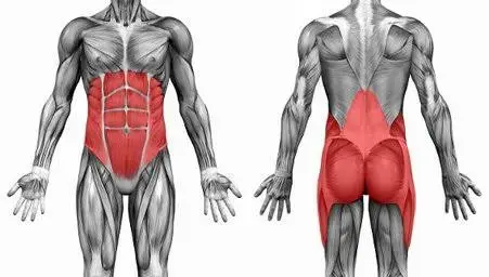
The pelvic floor, abdominal wall, back muscles, and diaphragm muscles are the main muscles that stabilize and regulate the pressure inside the trunk. These muscles make up the core muscles.
- Pelvic floor
A muscular sheet with a dome shape that divides the perineal area below from the pelvic cavity above is called the pelvic floor. The pelvic viscera—the uterus, the bladder, and the intestines—are enclosed in this cavity in females. The pelvic floor muscles serve the following primary purposes:
- To bolster the viscera of the abdomen and pelvis
- to keep one’s faeces and urine in check
- permits giving birth, vaginal sex, defecation, and sexual activity
Layer of muscle
Layer 1
- The Urogenital Triangle
Layer 2
- Urogenital Diaphragm Bulbocavernosus Ischiocavernosus Superficial transverse perineal Outside anal sphincter
- The urogenital diaphragm, which is also referred to as the triangular ligament, is a robust and muscular membrane that spans the anterior triangular portion of the pelvic outlet and lies between the symphysis pubis and ischial tuberosity. The pelvic diaphragm is superior to the urogenital diaphragm, which is external.
- Sphincter in the urethra
Layer 3
- Sphincter urethral (sphincter urethrae)
- Compressor urethrae Sphincter vaginalis urethral
- Perineal membrane, deep transverse
- The Pelvic Diaphragm
The inferior border of the abdominopelvic cavity is formed by the broad but thin muscular layer known as the pelvic diaphragm. It is made up of a wide, funnel-shaped sling of muscle and fascia that runs from one lateral sidewall to the other and from the symphysis pubis to the coccyx.
- Levator Ani Muscle (also known as iliococcygeus, pubovisceral, pubovaginalis, puboanalis, and puborectalis)
- Coccyges distinctions
- Internal obturator
- Arcus Tendinous fasciae pelvis Arcus Tendinous of Levator ani
2. Abdominal muscle
These four primary muscles come to mind when one thinks of abdominal muscles.
Transverses abdominis
Its primary functions include maintaining internal abdominal pressure and stabilizing the trunk.
Situated at the front of the pelvis, between the pubis and the ribs, is the rectus abdominis. This muscle has distinct protuberances or lumps that are referred to as “the six-pack” when it contracts. The rectus abdominis’ primary job is to transfer the body’s weight between the pelvis and the ribs.
External oblique muscles
The muscles on either side of the rectus abdominis are known as external oblique muscles. The trunk can twist, but only to the opposite side of the external oblique muscle that is contracting, thanks to the external oblique muscles. For example, the right external oblique contracts, causing the body to turn to the left side.
Internal oblique muscles
Just inside the hipbones are the internal oblique muscles, which are situated to the side of the rectus abdominis. Compared to the external oblique muscles, they function oppositely. For example, to twist the trunk to the left, the external oblique on the right side and the internal oblique on the left must contract simultaneously. Patients with persistent low back pain can strengthen their deep trunk muscles with core strength training. For the treatment of chronic low back pain, core strength training—which includes strengthening the deep back muscles—is more beneficial than traditional resistance training.
The function of abdominal muscle
The abdominal muscles are connected by either a common site of connection or by fascia. The muscles have distinct orientations of muscle fibres and act in all three planes during movements. Control of the abdominal muscles involves sometimes intricate movements. Normally, a single muscle works in unison with other muscles rather than in isolation. There are almost endless variations in physical activity, all controlled by the brain. such as During gait, the abdominal muscles cooperate to regulate the movement of the rib cage, pelvis, and spine. The upper and lower limbs counter-rotate, and the arm and leg move in opposite directions from one another. In a typical gait, there’s a point at which one side’s external obliques and rectus abdominis work eccentrically to slow down the anterior pelvic tilt brought on by that side’s extended hip.
To regulate the thoracic extension and rotation brought about by the extension of the shoulder, the RA and the external obliques on the opposing side collaborate eccentrically. Abdominal muscles are used in almost every action, including swimming, chess, golf club swinging, biking, running, and walking. Together with other core muscles, the abdominals contribute to the body’s stability and balance even when it is at rest.
3. Diaphragm
One important breathing muscle is the diaphragm, The “fibromuscular sheet” that divides the thorax from the abdomen has a dome-like form. It forms the roof of the abdomen as well as the floor of the thorax Because the liver is located on the right side, the left side is lower than the right. The “push” of the heart may also cause the left side to be partially located inferiorly.
Three separate muscle groups make up the muscular peripheral portion of the diaphragm:
- The xiphoid process gives rise to two fleshy slips that make up the sternal group. The inner surfaces of the cartilages and the surrounding areas of the six lower ribs are the source of the coastal group.
- The lumbar group is the source of the arcuate ligaments and the two crura, which are subsequently inserted into L1 and L2, and occasionally L3 as well.
- Very powerful aponeurosis tendinous ligaments, which lack bony attachments, comprise the central region of the diaphragm.
supply of motor nerves: The left phrenic nerve supplies the left hemidiaphragm. The right phrenic nerve supplies the right hemidiaphragm.
supply of sensory nerves: The peritoneum, which covers the diaphragm’s central surfaces, and the parietal pleura are innervated by the phrenic nerve. The diaphragm’s outer edge is innervated by the bottom six intercostal nerves. Large-diameter myelinated, small-diameter myelinated, and unmyelinated fibres make up the phrenic nerve. When the diaphragm contracts, the large-diameter fibres fire, but the small-diameter fibres. Sympathetic motor outflow is modulated by phrenic nerve activation. Phrenic afferents are responsible for somatosensation of the diaphragm and awaken people’s awareness of their breathing.
Strong Core vs Back Sprain
Strong core muscles help you stay balanced, avoid awkward movements, and shield your body from unintentional sprains or strains. Additionally, they greatly lower your chance of developing back pain by enabling your body to transfer force and stress through your muscles rather than your spine. Your back’s muscles support and align your vertebrae, as well as your spinal joints. To prevent back pain, abdominal muscles must maintain a neutral pelvic tilt and the appropriate curvature of the spine. The pressure inside your abdominal cavity rises when you contract your abdominal muscles. By doing this, you relieve pressure and weight from your spine.
When it comes to avoiding back pain, the other muscles in your core are also crucial. For instance, weak hip muscles impair balance, but strong gluteus muscles support your hip joint. The core muscles function as a unit to prevent disc issues and back injuries, which can result in severe pain and immobility.
Causes of the pulled muscle in the Lower Back
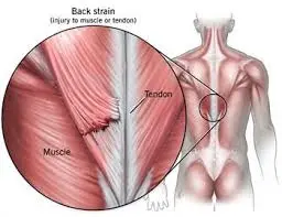
A sprain can result from overexerting or tearing the bands of tissue called ligaments that hold the spine’s vertebrae in place. A strain is a rip in one of the tendons that connect the muscles to the spinal column, or in the muscle itself. When someone stretches their muscles or muscle fibres farther than they should, they can get strained. Possible causes include:
- twisting overstretching lifting bulky items pushing and tugging activities like football or soccer
- Additional risk variables consist of:
- stooping the lower back excessively feeble abdominal or back muscles
- ill alignment and taut hamstrings
Additional factors that may be causing lower back pain on one or both sides are as follows:
- obesity-related slipped disc
- kidney infection
- endometriosis and kidney stones
Symptoms of the Pulled Muscle in the Lower Back
A person may experience a pop or tear as a lower back muscle twists or pulls as a result of an abrupt movement or injury.
The following are signs of a pulled lower back:
- diminished functionality and limited range of motion
- difficulty standing, bending, or walking straight; bruises and swelling; cramps or spasms in the muscles
- unexpected lower back pain
When to see the doctor if you suspect a pulled muscle in the lower back?
- See a doctor if the pain does not go away after 1-2 weeks.
- There are situations when someone needs to call 911 or visit the emergency room.
- When someone is injured, they hear a crack.
- The affected area of the back feels cold to the touch, discoloured, or numb.
- The pain is accompanied by a fever of 101°F (38.3°C).
- The person can no longer control their bowels or bladder.
- When someone has to urinate, it hurts and/or results in blood in the urine.
- There’s excruciating abdominal pain
Diagnosis of pulled muscle in the lower back
To identify a lumbar strain, a physician will probably conduct a physical examination. Should additional testing be necessary to determine the reason behind the lumbar strain, the physician may ask for:
- A CT scan or an X-ray
- an MRI examination
- A radionuclide bone scan to assess blood flow to the bone and cell activity, or an electromyogram to assess muscle and nerve function, may also be ordered.
Differential Diagnosis
Mechanical back pain
For some providers, mechanical back pain is a catch-all term, but with the right examination techniques, it can be a precise diagnosis. When joints experience mechanical back pain, it indicates that their mechanical behaviour is not optimal. Every bone on either side of a joint complex has an ideal physiological position. Traumatic forces, postural habits, the accumulation of lifting forces, scarring, ligamentous instability, sleeping in an awkward position, etc. can all affect this position. An imbalanced joint can cause abnormal strains on the surfaces of the joint, abnormal stresses on the joint capsules, and possibly even disrupt joint nutrition, all of which can trigger nociceptive pain receptors.
Back sprain
A sprain differs from a strain in that it is caused by an overloading of ligaments as opposed to overworked muscles. Ligaments are structures made of connective tissue that stabilize the skeleton by joining bones together. A sprain results in either partial or total ligament tearing. When repeated forces are avoided, ligaments typically heal over up to 12 weeks. Repetitive trauma can result in scarring that is difficult to heal and tearing of the healing tissue. Ligament injuries can cause a joint complex to become unstable, making it impossible for the joint surfaces to function together. This is particularly true if the ligament is entirely torn.
Back pain caused by discs
The intervertebral disc, a ligamentous ring, is the main joint connecting the vertebral bodies at different levels of the spine. It has few pain receptors when it’s not injured, but when it is, chemicals released as a result of the damage tend to cause more pain fibres to grow. The disc is prone to acute and chronic loading injuries as well as rotational injuries. Its nucleus can tear entirely through the rings of fibrocartilage or pierce through the layers of fibrous cartilage. Herniation will have its treatment plan. Tears allow chemicals and proteins that were kept out of the body’s immune system before birth to seep out, which triggers an inflammatory reaction.
Pinched nerve
When you overstretch or tear a portion of the muscle fibres, you get a pulled muscle. This may occur if you overuse the muscle or twist it excessively. You’ll likely experience pain, swelling, and tenderness in the affected area. You might even spot bruises or redness. A pinched nerve, also known as nerve compression, results from partial blockage of nerve impulses due to pressure in a particular area. A burning pain that radiates may be felt in the affected area.
A pinched nerve in your lower back may result from a pulled muscle, but a herniated disc in your spine can also be the cause of this. Visit a doctor straight away if you experience radiating pain that travels down your legs.
Spinal stenosis
When a person is diagnosed with spinal stenosis, their nerve roots are compressed due to either centrally encroaching bony arthritis or a narrowing of the openings between adjacent vertebrae. Due to increased nerve root pinching caused by the spine’s extension closing the foramina, or spaces between vertebrae, a person with spinal stenosis will frequently find it intolerable to stand upright. When flexion opens the foramina to a slight degree, decompressing the nerve root, the patient with spinal stenosis frequently assumes a forward bent posture that gets increasingly more pronounced. Patients with spinal stenosis will find it extremely difficult to descend stairs.
Post-laminectomy syndrome
Post-laminectomy syndrome, also known as failed back surgery syndrome (FBSS), is primarily caused by the growth of scar tissue that adheres to or compresses the nerve root. About 90% of disc surgeries are successful. Scarring is a normal part of the healing process following surgery, but in certain people, the healing process results in the generation of more scar tissue than is necessary. This causes the scar tissue to cross tissue layers and send out tendrils, which can tether nerve tissue that needs to be free to move or directly compress a nerve. Misdiagnosis and smoking, which affects the metabolism of bones and small blood vessels necessary for healing, are other causes of FBSS.
Arachnoiditis
The layers of connective tissue that envelop the brain and spinal cord are called meninges, and the arachnoid is their middle layer. It may result from an infection, such as meningitis caused by bacteria or viruses. It might happen following a bleed into the CSF.
Visceral-related back pain
Back pain that originates in the guts due to disease processes or mechanical factors is referred to as visceral back pain. There is a significant disparity. One possible cause is a growing aortic aneurysm-related back pain. It might be pain from an acute inflammatory response, like pancreatitis, or from a stomach ulcer that is eroding the stomach’s back wall. It may result from internal endometriosis scar tissue-related mechanical issues following abdominal or pelvic surgery, typically more than six months following the procedure.
Treatment of the pulled muscle in the lower back
There are a few ways to reduce pain and swelling if you do pull a lower back muscle.
Use heat or ice.
To minimize swelling, it’s a good idea to apply ice to your back straight away. But avoid putting an ice pack right against your skin. For ten to twenty minutes at a time, place it on the sore spot after wrapping it in a towel. A few days later, you can start using heat. Make sure you avoid sleeping with a heating pad and limit how long you leave it on to no more than 20 minutes at a time.
Anti-inflammatory drugs
Anti-inflammatories available over-the-counter (OTC), such as naproxen (Aleve) or ibuprofen (Advil), can help lessen swelling and inflammation, which will help ease pain. These drugs have a lot of potential side effects and should not be used for extended periods, even though they can be very effective. Make sure anti-inflammatories do not conflict with any of your current medications. Visit your pharmacy to find anti-inflammatories for kids. Over-the-counter medications can aid in the management of pain and edema. Take an ibuprofen or aspirin.
Massage
Massage can ease tense muscles and lessen pain. You can apply over-the-counter (OTC) creams that reduce pain in your skin.
Compression
By compressing the muscles, you can reduce swelling, which will help you manage your pain.
Brace
A back brace is probably necessary for your lower back to be effectively compressed. Take care not to overtighten it and take it off occasionally. Blood flow is essential for muscle healing.
Relax and rest
Bed rest may help you feel better, but it’s not advised unless you need it for short periods. Lie on your back on the floor with your knees bent, or on your side with a pillow beneath you.
After pulling a back muscle, you might find it helpful to reduce your activity for a few days, but staying in bed for longer than that can weaken your muscles. It’s preferable to build
Exercises of pulled muscle in the lower back
You can do a variety of exercises to aid in the healing of your lower back. They not only relieve any muscle spasms you may be experiencing, but they also strengthen your back, reducing the likelihood of further injury These are some simple stretches that you can do. Move into each position gradually, taking them one at a time. If any of these exacerbate your back pain, stop and consult a physician.
Twists
- With your legs fully extended on the forward side of you, sleep on your back or in a prone position.
- Cross your right leg over the left side of the body and slightly bend your right knee.
- Hold it so that your back feels as though it is gently stretching.
- After 20 seconds of holding, switch to the other side.
- Do this three times.
Knee pull
- On a level surface, bend at the knees so that they are beneath your hips and your hands are directly under your shoulders.
- Breathe out and allow your back to gently curve downward.
- Take a breath and raise your back.
- Give each position a ten-second hold.
- Ten times over, repeat.
Cat and cow pose
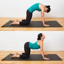
- On a level surface, bend at the knees so that they are beneath your hips and your hands are directly under your shoulders.
- Breathe out and slowly allow your back to bend in a downward direction.
- Take a breath and raise your back.
- Give each position a ten-second hold.
- Ten times over, repeat.
Back muscle stretch
Exercises that involve bringing the knees and chin to the chest while lying on one’s back can cause minor straining of the torso, shoulders, and neck muscles. By extending these muscles, you can reduce the risk of muscle strain and increase the flexibility of the spine. Stretch with thirty second hold.
Hip and glute muscle stretch
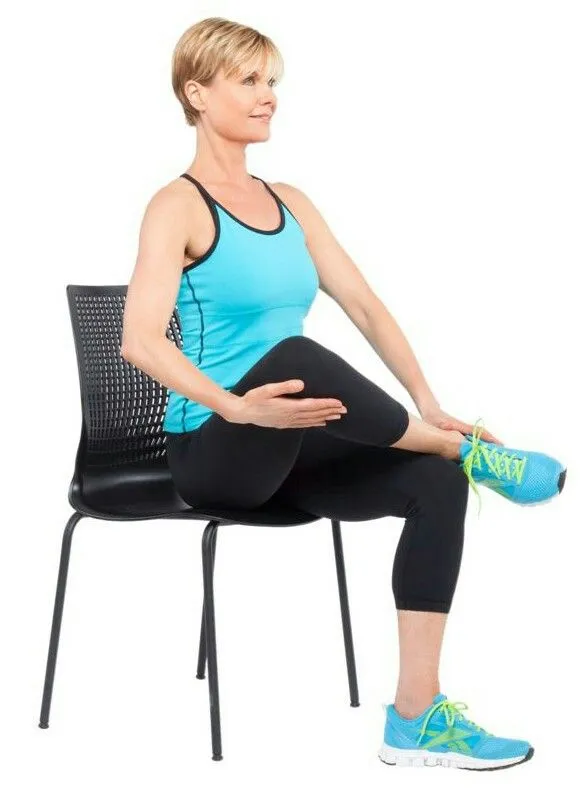
The gluteus, piriformis, and hip flexor muscles are the main targets of these stretches. By stretching these muscles, you can preserve a healthy range of motion and reduce lower body tension. Stretching the piriformis muscle, for instance, involves lying on one’s back and pulling one knee across the body and up to the chest, leaving the other leg flat. The buttocks or upper thighs should feel slightly pulled as a result. Stretch with thirty second hold.
Hamstring stretch
Stretches for the hamstrings are intended to gradually lengthen the muscles, reducing strain on the lower. The hamstring muscles run from the back of the thighs to the knee, starting in the pelvis. Stretching your hamstrings gently and with support can help protect your lower back.
Stretching should not cause pain; if it does, you should stop. Stretches should be held for 20 to 30 seconds, or until you feel the muscles starting to loosen up, and then performed five to ten times. Deep breathing eases tense muscles and facilitates stretching.
Strengthening exercises for the Pulled muscle in the lower back
Exercises to build or enhance the strength of the muscles in the lower back and core are numerous. Pilates, yoga, and tai chi are frequently advised practices, in addition to working with a physical therapist or other healthcare professionals who implement a personalized exercise regimen.
Transverses Abdominal Muscle Strengthening (Abdominal Exercise)
To strengthen the abdominal muscles, many people consider doing sit-ups or crunches. While some people think “six-pack abs” look good, working the Transverses Abdominis (TVA) through abdominal exercises is more crucial to achieving spinal stability. Maintaining a neutral lumbar spine is crucial when retraining the TVA (don’t try pushing the back into the floor). Strengthening the back in a flexed or extended position makes less sense because the back is typically in a neutral spine position.
Bend your knees while lying on your back. Shoulder width should separate the knees and feet. Keeping the spine neutral, draw the belly button in the direction of the spine. When you release your breath, pretend to reach for the ceiling. Next, lift your head and shoulders off the ground until your shoulder blades barely touch the ground. As soon as you’re back in the air, take another breath and repeat. Continue until you become fatigued or it becomes impossible to keep your spine neutral. Hold for one or two seconds. Repeat 4-5 days a week, one time per day, until you become exhausted.
Stabilization exercise for the Pulled muscle in the lower back
The range of exercises in the lumbar stabilization exercise programme progresses from basic to more difficult.
- From motionless (lying) to active (standing or leaping)
- Moving from defying gravity to defying another external force
- Transitioning from dependable to erratic motions
- From the individual parts of a movement to the movement’s entire range of motion
The neutral spine position is upheld at all times. In most cases, mastering the neutral spine technique during the current exercise is a prerequisite for moving on to the next one. The exercise therapist or physical therapist is qualified to assist the patient in picking up the appropriate form.
Aerobic exercise for the Pulled muscle in the lower back
Aerobic exercise can be helpful for recovery and rehabilitation following a back muscle injury. Healthy blood flow, which is promoted by aerobic exercise, carries nutrients and oxygen to wounded muscles throughout the body, promoting tissue healing.
Low-impact exercise is a fantastic way to get more exercise while minimizing back pain because it raises heart rate without jarring the body. Low-impact aerobic workouts include the following:
Walk for exercise
Walking quickly and vigorously can increase heart rate without overly straining the lower back.
Stationary cycling
The benefits of riding a stationary bike include increased flexibility and strength in the legs, hips, and back without the strain of riding on uneven ground outside. Perhaps a regular stationary bike or a recumbent bike would be better.
Elliptical training
An elliptical machine can be used to simulate running without having to worry about your feet striking the floor. With the use of elliptical machines, users can increase their heart rate while strengthening their lower back, core, hips, and buttocks muscles.
Swimming, water aerobics, or water therapy
Even if you have back pain, working out in a pool can still be comfortable and low risk of injury. In addition to providing comfortable movement with minimal effect on the spine, the buoyancy and resistance of swimming help to strengthen all of the body’s muscles, particularly the back and core. A lot of pools are maintained at a warm temperature, which is frequently comforting for strained lower back muscles.
McKenzie’s approach is to pull a muscle in the lower back
New Zealand physical therapist Robin McKenzie created the McKenzie Method in the 1960s. In his practice, he observed that in some patients, extending the spine could result in significant pain relief and enable them to resume their regular daily activities.
By shifting the patient’s pain from the extremities (arm or leg) to the back, physical therapy and exercises that stretch the spine can help “centralise” their pain according to the McKenzie method. Back pain is typically more tolerable than arm or leg pain, and the idea behind this approach is that by treating the source of the pain instead of just the symptoms, back pain can be effectively managed. The technique itself focuses on categorizing spinal diseases or disorders according to their symptoms and how they react to particular preliminary evaluation techniques. The patient is given instructions on specific exercises to gradually centralize or eliminate the pain based on the classification. Among the classifications are:
- Postural syndromes: persistent strain on soft tissues while sustaining specific positions or postures can result in back pain.
- Derangement syndromes: back pain that varies with repeated motion due to a shift in the vertebrae surrounding a disc as a result of the disc’s fluid nucleus shifting in place.
- Dysfunction syndromes are characterized by sporadic back pain and restricted range of motion brought on by the presence of shortened scar tissue; back pain is brought on by stress on these tissues.
Posture management of the Pulled muscle in the lower back
- Pose correctly to avoid getting hurt again.
- Bend at the knees and hips when you move. Don’t twist or bend at the waist.
- Keep the things near to your body while object lifting. Never attempt to lift more than you are capable of.
- Maintain a supported lower back when seated. When necessary, use a rolled-up towel.
Duration of recovery of the pulled muscle in the lower back
The majority of patients with lower back sprains and strains should heal in two weeks, and over 90% do so in a month, according to the American Association of Neurological Surgeons (AANS).
For eight weeks, a person should refrain from strenuous exercise to lessen the chance of further back damage.
Live with a pulled muscle in the lower back
- Swelling is decreased by cold. Heat and cold both help to lessen pain. Towels should be placed between your body and the heat or ice to protect your skin.
- Use an ice pack for 15 to 20 minutes every day for the first few days.
- Try applying heat for 15 minutes at a time to relieve pain after the first few days. Never use a heating pad to fall asleep.
- Over-the-counter medications can aid in the management of pain and edema. Take an ibuprofen or aspirin.
Prevention of the pulled muscle in the lower back
You can take precautionary measures as well as exercises that help strengthen your lower back to avoid straining it. Among them are:
- exercises for strengthening and extending
- strolling, swimming, or engaging in other mild cardiovascular exercises
- reducing weight strengthening your posture when standing and sitting, taking precautions to prevent falls, and donning low-heeled, supportive footwear
- reclining on your side with your knees bent up on a comfortable mattress
Take away note for the pulled muscle in the lower back
Although most people will experience lower back pain at some point, these wounds typically recover in a few days. Using over-the-counter topical creams, ice packs, and gentle stretching can all help hasten the healing process. Frequent back injury prevention can be achieved by strengthening your back muscles through exercise.
See a doctor, though, if you have other symptoms like weakness and fever, tingling in your legs and feet, or if you have pulled a muscle in your lower back.
Summary
It can hurt to have a pulled lower back muscle. However, most strains recover in a few weeks with the proper at-home care. If someone hears a crack at the scene of the injury, gets a fever, or becomes incontinent afterward, they should visit a doctor.
A person is more susceptible to sprains and strains if they have weak muscles or carry excess weight. It is crucial to exercise after warming up and to handle heavy objects with caution.
FAQs
typically disappear in four to six weeks.
Avoid standing still for too long. There’s a chance that your muscles will weaken and become more painful and stiff.
A strain is an injury to a muscle or tendon caused by tissue stretching or tearing.
To treat a thrown-out back and back muscle strain properly, pain management techniques like resting and limiting walking are necessary.
References
- TeachMeAnatomy. (2020, October 29). The superficial back muscles – attachments – actions – TeachMeAnatomy. https://teachmeanatomy.info/back/muscles/superficial/
- Back muscles. (n.d.). Physiopedia. https://www.physio-pedia.com/Back_Muscles
- Abdominal muscles. (n.d.). Physiopedia. https://www.physio-pedia.com/Abdominal_Muscles
- Eagle, R. (2023, March 15). How to treat and prevent a pulled muscle in the lower back. https://www.medicalnewstoday.com/articles/pulled-muscle-in-lower-back#summary
- Mooney, V., MD. (n.d.). What is the McKenzie Method for Back Pain and Neck Pain? Spine-health. https://www.spine-health.com/wellness/exercise/what-mckenzie-method-back-pain-and-neck-pain
- Cole, A., MD. (n.d.). Lumbar spine stabilization exercises. Spine-health. https://www.spine-health.com/wellness/exercise/lumbar-spine-stabilization-exercises
- Articles. (n.d.). https://www.cedars-sinai.org/health-library/diseases-and-conditions/l/lumbar-strain.html
- TeachMeAnatomy. (2023b, January 19). The intrinsic back muscles – attachments – actions – TeachMeAnatomy. https://teachmeanatomy.info/back/muscles/intrinsic/
- Morse, R. G. (2019, May 7). What you need to know about treating lower back muscles. Healthline. https://www.healthline.com/health/pulled-muscle-in-lower-back#summary
- Bonaventure Ngu, MD. (n.d.). Building a Strong Core is Your Best Defense Against Back Pain: Bonaventure Ngu, MD: Orthopaedic Spine Surgeon. https://www.premierspineinstitute.com/blog/building-a-strong-core-is-your-best-defense-against-back-pain
- TeachMeAnatomy. (2023, January 5). The intermediate back muscles – attachments – actions – TeachMeAnatomy. https://teachmeanatomy.info/back/muscles/intermediate/