Muscles Of Back Of Leg
Table of Contents
Introduction:
Anatomically, the leg is defined as the part of the lower limb below the knee. It consists of three-compartment, posterior, anterior, and lateral compartments. In accordance, the muscles of the leg are organized into three different groups:
The Anterior group(dorsiflexor group), contains the tibialis anterior, extensor digitorum longus, fibularis tertius, and extensor hallucis longus muscles.
The Posterior group(plantar flexor group), consists of a superficial layer comprised of the gastrocnemius, plantaris, and soleus, and a deep layer comprised of the tibialis posterior, flexor hallucis longus, popliteus, and flexor digitorum longus muscles.
The lateral group (fibular group), consists of fibularis longus and fibularis Brevis muscles.
The muscles of the back of the leg are classified into two groups:
- Superficial muscles of Muscles of Back of leg
- deep muscles of Back of leg
Superficial muscles include Gastrocnemius muscle, Soleus, and Plantaris muscles
Deep muscles include Popliteus, Flexor hallucis longus, Flexor digitorum longus, and Tibialis posterior muscles
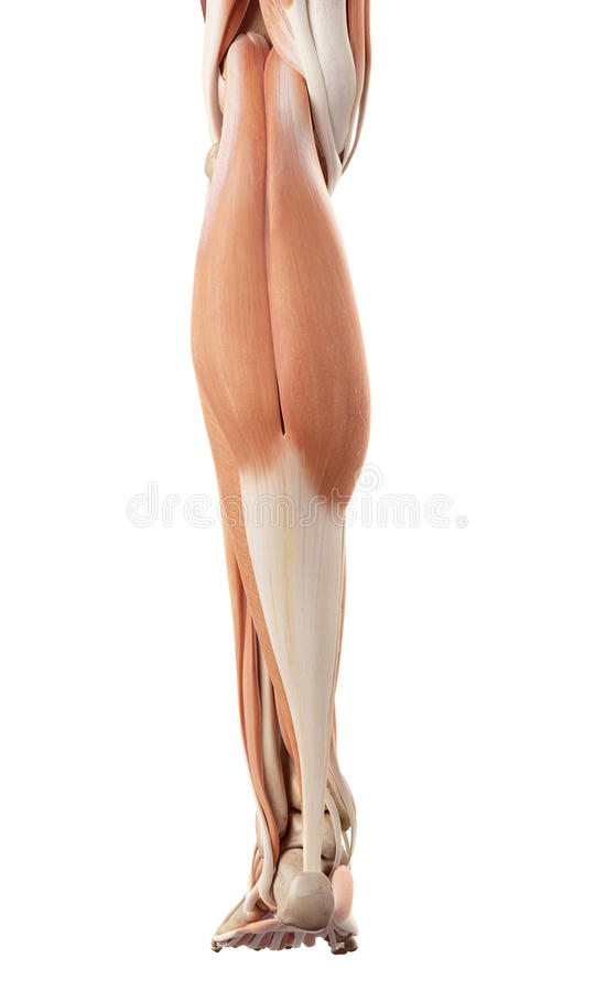
Superficial Muscles Of Back Of The Leg
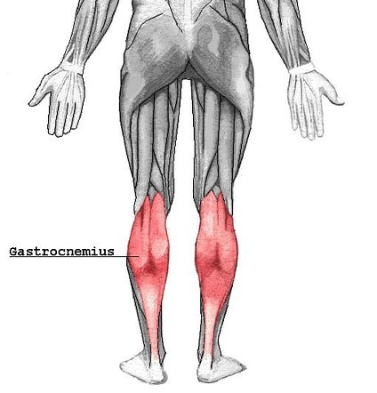
Gastrocnemius muscle
Origin:
The medial head of the Gastrocnemius muscle is larger than the lateral head of the Gastrocnemius muscle
It arises from a broad flat tendon from:
The posterosuperior depression on the medial condyle of the femur bone, behind the adductor tubercle
The adjoining raised area on the popliteal surface of the femur bone
The capsule of the knee joint
The lateral head of the Gastrocnemius muscle has arisen from a broad flat tendon from:
The lateral surface of the lateral condyle of the femur bone
The lateral supracondylar line
The capsule of the knee joint
Insertion:
The tendon of this muscle fuses with the tendon of the soleus to form the tendocalcaneus or tendoachillies, which is inserted into the middle one-third of the posterior surface of the calcaneum
Nerve supply:
Tibial nerve (S1, S2)
Blood supply:
Both medial heads of gastrocnemius muscles and lateral heads of gastrocnemius muscles blood supply are the lateral and medial sural arteries, which are direct branches of the popliteal artery.
Action:
Talocrural joint: Foot plantar flexion
Knee joint: Leg flexion
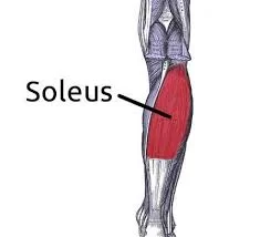
Soleus muscle
Origin:
It has a dome-shaped origin from:
The fibula: the back of the head, and the upper one-fourth of the posterior surface of the shaft.
The tibia: Soleal line and middle one-third of the medial border of the shaft
The tendinous soleal arch stretches between the tibia and the fibula
Insertion:
The posterior surface of the calcaneus (via calcaneal tendon)
Nerve supply:
The nerve supply of the Soleus muscles is the Tibial nerve (S1, S2)
Blood supply:
The arterial supply of the Soleus muscles is the Posterior tibial artery and vein
Action:
Talocrural joint: Foot plantar flexion
Plantaris muscle:
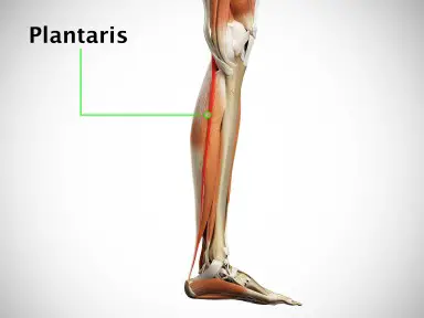
Origin:
Plantaris muscles originate from the lower part of the lateral supracondylar line of the femur
oblique popliteal ligament of the knee
Insertion:
The tendon is thin and long. It lies between the gastrocnemius and the soleus, crossing from the lateral to the medial side
It is inserted on the posterior surface of the calcaneus (via calcaneal tendon). Planter aponeurosis is the estranged part of the plantaris
Nerve supply:
The nerve supply of the Plantaris muscle is the tibial nerve (S1, S2)
Blood supply:
Superficially: lateral sural and popliteal arteries
Deeply: superior lateral genicular artery
Action:
Talocrural joint: foot plantar flexion
Knee joint: knee flexion
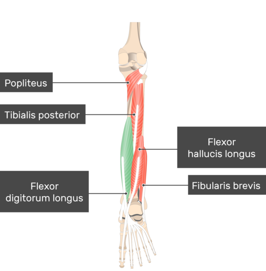
Deep Muscles Of Back Of The Leg
Popliteus muscle
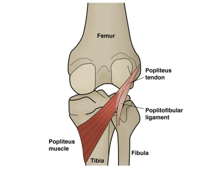
Origin:
Popliteus muscles originate from,
The lateral surface of the lateral condyle of the femur bone,
Origin is intracapsular
And Outer margin of the lateral meniscus of the knee joint
Insertion:
The posterior surface of shaft of tibia above soleal line
Nerve supply:
The nerve supply of the popliteus muscles is the tibial nerve (L5-S2)
Blood supply:
The popliteus receives arterial blood supply mainly from branches of the popliteal artery, namely the inferior medial genicular arteries and lateral genicular arteries.
Action:
Unlocks knee joint by lateral rotation of the femur on the tibia to flexion
Flexor hallucis longus muscle
Origin:
The Flexor hallucis longus muscles originate from the Lower three-fourths of the Posterior surface of the fibula and the interosseous membrane
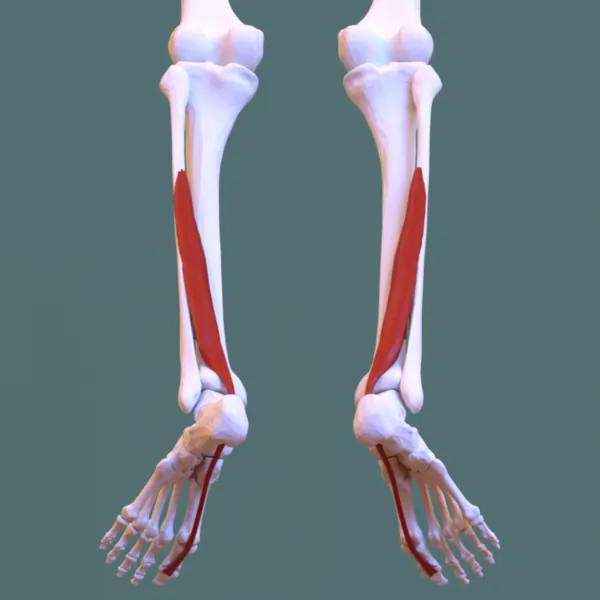
Insertion:
The Flexor hallucis longus muscles are inserted on the plantar surface of the base of the distal phalanx of the big toe
Nerve supply:
The nerve supply of the Flexor hallucis longus muscle is the Tibial nerve (S2, S3)
Blood supply:
Flexor hallucis longus muscles receive arterial supply from the branches of the posterior tibial and fibular arteries
Action:
Flexes distal phalanx of the big toe, plantar flexor of ankle joint, supports the medial longitudinal arch of the foot
Flexor digitorum longus
Origin:
the Flexor digitorum longus muscles originate from the Upper two-thirds of the medial part of the posterior surface of the tibia below the soleal line
Insertion:
the Flexor digitorum longus muscles are inserted on the Bases of distal phalanges of the shaft of lateral four toes. The muscle ends in a tendon divided into four slips, one for each of the lateral four toes. Each slip is attached to the plantar surface of the distal phalanx of the concerned
Nerve supply:
The Flexor digitorum longus muscle’s nerve supply is the tibial nerve (S1, S2)
Blood supply:
The arterial supply of the Flexor digitorum longus muscle is the posterior tibial artery
Action:
Flexes distal phalanges, plantar flexor of the ankle joint supports medial and lateral longitudinal arches of the foot
Tibialis posterior muscle
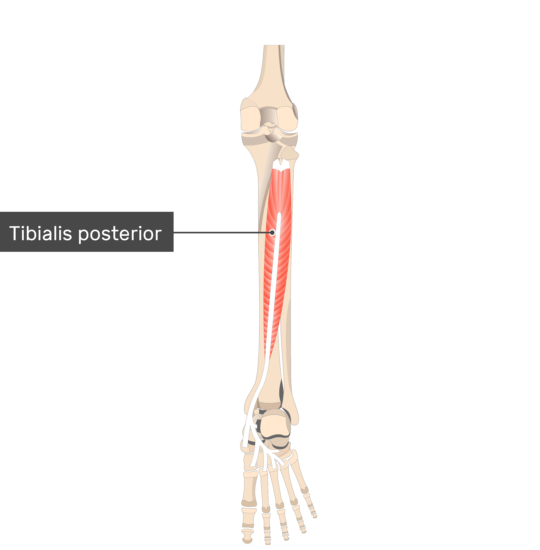
Origin:
The Tibialis posterior muscles originate from the upper two-thirds of the lateral posterior surface of the tibia below the soleal line. The posterior surface of the fibula in front of the medial crest and the posterior surface of the Interosseous membrane
Insertion:
The tibialis posterior muscles are inserted on the tuberosity of the navicular bone and other tarsal bones except for the talus. Insertion is extended into 2nd,3rd and4th metatarsal bones at their bases
Nerve supply:
The nerve supply of the Tibialis posterior muscle is the tibial nerve (L4, L5)
Blood supply:
The vascular supply to the tibialis posterior muscles is predominantly via the posterior tibial artery, which arises from the popliteal artery.
Action:
Plantar flexion of ankle joint; inverts foot at Subtalar joint; Supports the medial longitudinal arch of the foot
Clinical significance:
Medical conditions that result in calf swelling, among other symptoms, include deep vein thrombosis (DVT), compartment syndrome, Achilles tendon rupture, and varicose veins.
Idiopathic leg cramps are most common and typically affect the calf muscles at night. Edema is also common and, in many cases, idiopathic. Wearing compression garments helped to reduce edema and the pain related to edema. A small study of runners found that wearing knee-high compression stockings while running can significantly improve performance.
Most leg pain is caused by wear and tear, overuse, or injuries in joints or bones or in muscles, ligaments, tendons, or other soft tissues. Some types of leg pain may be traced to problems in your lower spine. Leg pain can also be caused by blood clots, varicose veins, or poor circulation in the lower limbs.