Tibial Tuberosity Anatomy
Table of Contents
Introduction
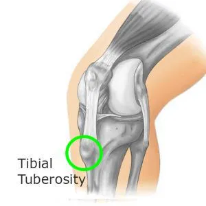
- On the anterior aspect of the proximal tibia, there is a big, broad protrusion known as the tibial tuberosity.
- It is easily palpable as the protrusion that is situated right below the patella. The infrapatellar bursa, which is Subcutaneous, lies beneath the tibial tuberosity.
- The patellar ligament, commonly known as the common quadriceps tendon, has its inferior insertion at the Tibial tuberosity.
- English terminology: tibial tuberosity Tuberositas tibiae in Latin.Meaning and purpose Large bony projection On the proximal tibia’s front side. acts as the patellar Ligament’s attachment point.
Relevant Anatomy
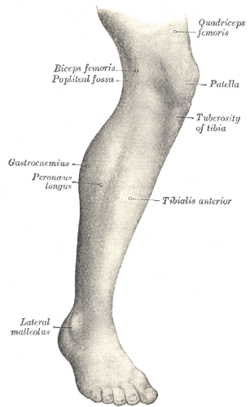
- The tuberosity of the tibia, also known as the tibial tuberosity or tibial tubercle, is an elevation on the Proximal, anterior part of the tibia that is located just below the point where the anterior surfaces of the Lateral and medial tibial condyles meet.
Structure of Tibial Tuberosity
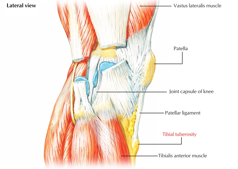
- The patellar ligament, which attaches to the patella from where the suprapatellar ligament forms the distal Tendon of the quadriceps femoris muscles, is attached to the tibial tuberosity.
- The quadriceps muscles Consist of the 4 group muscle which are rectus femoris, vastus lateralis, vastus medialis, and vastus intermedius. The femoral nerve supplies the innervation for these quadriceps muscles. As a result, the tibial tuberosity serves as the Terminal portion of a vast structure that extends the knee joint and keeps it from collapsing when the foot Hits the ground. The patella, the tibial tuberosity, and the two ligaments are all surface-level, easily Perceptible structures.
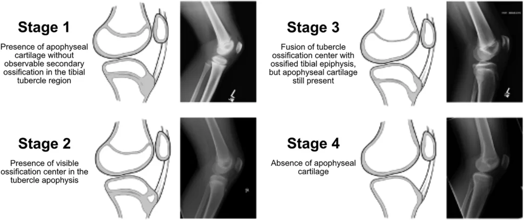
Stages
Fractures
- Teenagers are most frequently affected by tibial tuberosity fractures, which are uncommon fractures. Extreme knee extensor contraction during running and jumping activities might cause tuberosity apophysis avulsion fractures. If the fragment is not moved from its usual position on the tibia, only a cast is necessary. Surgery, however, is required if the fracture fragment is displaced in order to restore normal function.
- See also Osgood-Schlatter disease or deep infrapatellar bursitis can cause tibial tuberosity tenderness. In an adolescent with open growth plates or what is left of Osgood-Schlatter’s in adults, a bony protrusion on the tibial tuberosity may be the result of continuous Osgood-Schlatter’s irritation.
FAQ
Hyaline cartilage grows out and fibrovascular tissue grows in at the same time as the tibial tuberosity develops. This hyaline cartilage protrusion gradually moves distally during the fetal period until it is located next to the anterior part of the tibial metaphysis.
Where muscles and connective tissues attach, there is a modest protrusion called tuberosity. It performs comparable duties as a trochanter. The tibial tuberosity, deltoid tuberosity, and ischial tuberosity are a few examples. A tiny, spherical protrusion where connective tissues attach is called a tubercle.
The patellar tendon joins the quadriceps muscles (also known as quads) to the leg at the tibial tubercle, a bony protrusion on the top of the shin. A break or crack here is known as a tibial tubercle fracture, and it typically results from the patellar tendon pushing a portion of the bone away.
In healthy knees, the average TT-TG distance was 13 mm, but in pathological knees, it was 16.4 mm. There was no threshold value between normal and pathologic knees as a result of the measurement variability between normal and pathologic knees varying between 5 and 15 mm, respectively.
The patellar tendon links to the tibial tuberosity, a bulge on top of the tibia (shinbone). Tendons link bones and muscles. Over the patella (kneecap), the patellar tendon extends. The big quadriceps muscle on the front of the thigh is joined to the tibial tuberosity via the patellar tendon.
The anterior aspect of the proximal shaft of the tibia is home to the palpable bony protrusion known as the tibial tuberosity. The soleal line crosses the proximal third of the tibia on the posterior surface of the bone diagonally in a distal-to-medial manner.