
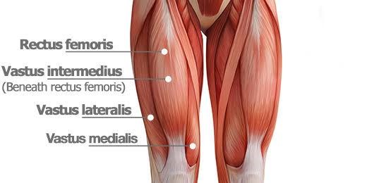
Quadriceps Muscles: Rectus femoris, Vastus Lateralis, Vastus Medialis, Vastus intermedius
Table of Contents
Introduction :
- The Quadriceps femoris is the most voluminous muscle in the human body.
- The Quadriceps femoris group of muscles is located in the anterior compartment of the thigh.
- The quadriceps femoris is hip flexors and knee extensors.
- It contains four individual muscles; three Vastus muscles and one rectus femoris. The Quadriceps femoris group of muscles form the main bulk of the thigh, and collectively they are one of the most powerful muscles in the body.
The four 4 sub-components are:
- Rectus femoris
- Vastus Lateralis
- Vastus Medialis
- Vastus Intermedius
Rectus femoris :
Origin :

The rectus femoris is a fusiform muscle that contains two heads. It originates from two sites on the ilium; the anterior inferior iliac spine (AIIS)(straight head) and supraacetabular groove (reflected head). The two heads unite into a common muscle belly that courses down the thigh in an almost vertical fashion, covering the anterior aspect of the thigh.
Insertion:
The rectus femoris muscle fibers converge towards a thick tendon that inserts into the base of the patella. Occasionally, the rectus femoris muscles can have a third head that originates from the iliofemoral ligament.
Relations
The proximal part of the rectus femoris muscle lies deep in Tensor Fasciae Latae(TFL), sartorius, and Iliacus muscles. All the contents of the anterior compartment of the thigh lie deep in the rectus femoris. These include the capsule of the hip joint, Vastus Intermedius, anterior margins of Vastus Lateralis and Vastus Intermedius muscles, lateral circumflex femoral artery, and some branches of the femoral nerve.
Nerve supply :
Rectus Femoris is innervated by the femoral nerve, originating from the lumbar nerve L2, L3, and L4 nerve roots
Blood supply :
The rectus femoris muscle is supplied by the artery of the quadriceps, which can stem from three sources; femoral, deep femoral, or lateral circumflex femoral arteries. The lateral circumflex femoral and superficial circumflex iliac arteries also contribute to the blood supply of the rectus femoris, but to a lesser extent.
Function :
Hip flexion
Rectus Femoris acts with iliopsoas to produce hip flexion especially when the knee is flexed.
During gait, as a hip flexor, it acts with the iliopsoas in the “Toe off” phase.
Knee extension
Vastus Lateralis :

The Vastus Lateralis is localized on the lateral side of the thigh. This is the largest of the quadriceps muscle that includes: the rectus femoris, Vastus Intermedius, and Vastus Medialis. the quadriceps muscles act on the hip and knee joints together to promote movement as well as strength and stability. They provide power for and absorb the impact of daily activities like walking, running, and jumping.
Origin :
The Vastus Lateralis muscles are originated from the Upper inter-trochanteric line, the base of the greater trochanter, lateral linea Aspera, lateral supracondylar ridge, and lateral intermuscular septum
Insertion :
Lateral quadriceps tendon which attached to the tibial tubercle.
Nerve Supply :
The nerve supply of the Vastus Lateralis is the Posterior division of the femoral nerve (L3,4)
Blood Supply :
Lateral circumflex femoral artery
Action :
Extension of the knee
Vastus Medialis :
Description :
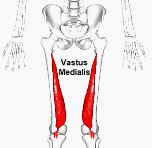
This muscle is one of the four muscles that make up the group of quadriceps muscles. Vastus Medialis muscle originates from the upper part of the femoral shaft and inserts as a flattened tendon into the quadriceps femoris tendon, which inserts into the upper part of the patella.
Origin :
This muscle originates from the lower part of the intertrochanteric line, along the spiral line to the medial lip of the linea Aspera, the medial intermuscular septum, and the aponeurosis of the adductor Magnus.
Insertion :
Vastus Medialis inserted Into the medial side of the quadriceps tendon, joining with rectus femoris and the other quadriceps muscles, enveloping the patella, then by the patellar ligament into the tibial tuberosity.
Nerve supply :
A branch from the posterior division of the femoral nerve, derived from L2, L3 andL4.
Blood Supply :
The blood supply of the Vastus Medialis is the Femoral artery and branches from the Profunda femoris artery.
Action :
Extention of the knee joint
Vastus Medialis also contributes to the correct tracking of the patella.
Vastus Intermedius :
Description :
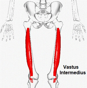
These muscles are localized in the center, under the Rectus femoris muscles in the anterior compartment of the thigh, and on each side of the Vastus Medialis and Vastus Lateralis respectively. The Vastus Intermedius muscle is one of the four muscles that make up the quadriceps femoris muscle. The tensor of the Vastus Intermedius is a new muscle that is part of the Quadriceps femoris.
Origin :
Vastus Intermedius muscles Originated from the upper two-thirds of anterior and lateral surfaces of the femur and the intermuscular septum.
Insertion :
Quadriceps tendon to base of the patella and onto tibial tuberosity via the patellar ligament.
Nerve supply :
The nerve supply of the Vastus Intermedius muscle is innervated by a branch of the Femoral nerve, originating from lumbar L2, L3, and L4 nerve roots.
Blood supply :
The blood supply of the Vastus Intermedius muscle is descending branch of the lateral circumference femoral artery.
Action :
Extension of the knee.
Quadriceps muscle strain
Definition :
A quadriceps muscle strain is an acute tearing injury of the quadriceps muscles. This injury is usually due to a stretch of the muscle, often at the same time as a forceful contraction or repetitive functional overloading of muscles.
Causes:
traumatic
The quadriceps are particularly active while jumping, hopping, or kicking. Too much tension may cause the muscle fibers to tear. This can be the result of either excessive force or repetitive movement.
There are generally three mechanisms of injury for a quadriceps tear:
- Sudden deceleration of the leg during kicking
- violent contraction of the quadriceps during sprinting and
- the rapid deceleration of an overstretched muscle during a quick change of direction.
Signs and Symptoms :
Symptoms :
- Pain
- Difficulty bending
- Difficulty in straightening the knee
- Leg weakness
- Sharp pain when running, jumping, or kicking
Sign :
- Swelling
- Range of motion decrease
- Muscle strength reduced
- Skin temperature increased
- ecchymosis
- Sore end-feel
Investigation :
There are several types of Investigation that can be used for muscle strains:
Radiographs
Ultrasound
Magnetic resonance imaging (MRI)
Treatment :
Medical Management
The use of nonsteroidal anti-inflammatory drugs(NSAIDs)
Surgical intervention may be necessary if there is a complete quadriceps muscle tear.
Indications for Surgery
large intramuscular hematoma,
a complete quadriceps tear
muscle tear with few or no agonist muscles, or
a partial (II degree) tear if more than half of the muscle belly is torn.
Physiotherapy Treatment :
Rice Therapy
The rest of the therapy during the healing process is based on the RICE therapy, which includes:
Rest,
Ice treatment
Compression
Elevation
Ankle toe movement :
this exercise is used to maintain and improve mobility of the Lower limb.
Isometric exercise of quadriceps muscles :
The position of the patient is supine lying. Asked the patient to contract the quadriceps muscles(by using the therapist’s fist or folded towel under the knee). Initially, the repetition is low then gradually increased. The exercise should be pain-free.
Straight leg raises :
Bend the knee of your normal lower limb at a 90-degree angle, planting the foot flatly on the floor. Stabilize the muscles on your straight leg by contracting your quadriceps muscles (the group of muscles on the front of your thigh). Inhaling slowly, lift the leg straight about six inches off the ground.
Strengthening Exercise of quadriceps muscles
46 Comments