Lateral pterygoid Muscle
Table of Contents
Introduction
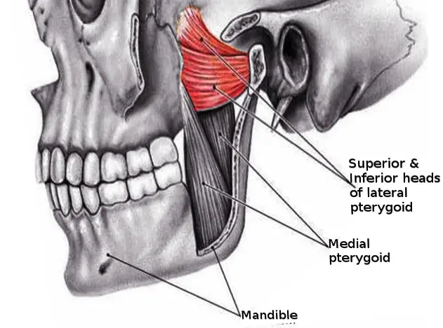
The lateral pterygoid muscle has two heads the upper head and the lower head.
The lateral pterygoid is the fan-shaped muscle located laterally in the infratemporal fossa of the skull. It is one of the four mastication muscles, along with the medial pterygoid muscle, temporalis, and masseter muscles.
All these mastication muscles act upon the temporomandibular joint (TMJ) to enable chewing (mastication) and biting. The lateral pterygoid muscle contributes to this function by protruding and depressing the mandible when contracting bilaterally, and by rotating the mandible when contracting unilaterally.
The lateral pterygoid muscle is supplied by pterygoid branches of the maxillary artery, and the lateral pterygoid nerve (from the mandibular nerve, CN V3).
Origin of Lateral pterygoid Muscle
Both heads of the lateral pterygoid muscles arise from the sphenoid bone.
The upper head is small. its origin is from the infratemporal surface and crest of the greater wing of the sphenoid bone.
The lower head is large. its origin is from the lateral pterygoid plate.
Insertion of Lateral pterygoid Muscle
The lateral pterygoid muscle fibers run backward and laterally and converge to be inserted into the pterygoid fovea on the anterior surface of the neck of the mandible and the anterior margin of the articular disc and temporomandibular joint capsule.
Nerve supply
The nerve supply of The lateral pterygoid muscle is the branches from the anterior division of the mandibular nerve (CN V3)
Blood supply
The blood supply of the lateral pterygoid is from the pterygoid branches of the maxillary artery and the ascending palatine branch of the facial artery.
Action
Depression of the mandible to open the mouth,(with suprahyoid muscle)
The lateral and medial pterygoid muscles of both sides act together to protrude the mandible.
The medial pterygoid muscle and lateral pterygoid muscles of both sides contract alternately to produce side-to-side movements of the mandible(as in chewing).
Clinical significance
The lateral pterygoid muscle can be involved in temporomandibular joint dysfunction.
Temporomandibular joint dysfunction (TMD, TMJD) is an umbrella term covering pain and dysfunction of the mastication muscles and the temporomandibular joints. The most important symptoms are pain, followed by restricted mandibular movement, and noises from the temporomandibular joints (TMJ) during jaw movement. Although TMD is not life-threatening, it can be detrimental to the quality of life; this is because the symptoms can become chronic and difficult to do management.
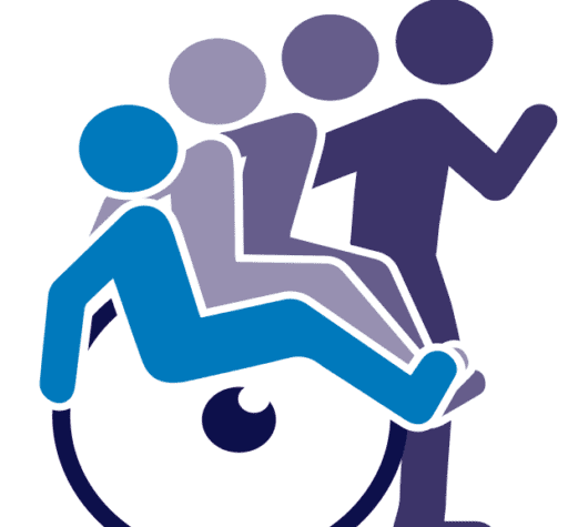
Exercise for the lateral pterygoid muscle
Stretching exercise for the lateral pterygoid muscle
Exercises to stretch the lateral pterygoid muscles can mimic the actual lateral movements of the lateral pterygoid muscles. Sit upright in a firm chair. Open your jaw 1 inch. Keep your mouth shut and slowly move your jaw as far as possible to your left without causing any pain. Hold for 10 seconds. Slowly return your jaw to the center position. then Relax. Repeat 10 times to the left, then do this exercise 10 times to the right.
Open your jaw approximately one inch. Then slowly move your jaw from one side to the other side without opening your jaw any further. If you have difficulty maintaining a one-inch gap you can clench a pen or pencil between your teeth while you perform the side-to-side movement of the jaw.