Cricoarytenoid Posterior Muscles
Table of Contents
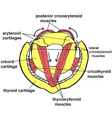
Cricoarytenoid Posterior Muscles Anatomy
The posterior cricoarytenoid muscle is a crucial component of the laryngeal musculature, playing a vital role in vocal function and respiration.
Situated within the larynx, this paired muscle originates from the posterior surface of the cricoid cartilage and inserts onto the muscular process of the arytenoid cartilage.
Its primary function is to abduct or open the vocal folds, allowing for the passage of air during inhalation and contributing to the production of sound during phonation. As one of the intrinsic muscles of the larynx, the posterior cricoarytenoid muscle is instrumental in modulating vocal pitch and controlling airflow, making it essential for various aspects of speech and respiratory physiology.
Origin:
posterior part of the cricoid.
Insertion:
muscular process of the arytenoid cartilage.
Nerve supply:
The posterior cricoarytenoid muscles are supplied by the recurrent laryngeal branch of the vagus nerve.
Blood Supply
The superior and inferior thyroid arteries’ laryngeal branches supply the posterior cricoarytenoid muscle. These are branches of the subclavian and external carotid arteries, respectively.
The internal jugular vein receives the venous blood after it has already passed through the superior and inferior laryngeal veins.
Function:
The posterior cricoarytenoid muscles are the only muscles to open the vocal cords. By rotating the arytenoid cartilages laterally, these muscles abduct the vocal cords and thereby open the rima glottidis.
Action:
abducts and laterally rotates the cartilage, pulling the vocal ligaments away from the midline and forward and so opening the rima glottidis.
This helps the other intrinsic muscles extend the vocal cords and splits the vocal cords, allowing air to travel through during inspiration and expiration. Because of this, the posterior cricoarytenoid muscle is the most crucial muscle in the larynx during breathing. Since this muscle is the sole one that can open the glottis, paralysis of this muscle can result in hypoxia and even death.
On the other hand, it is unclear exactly what part the posterior cricoarytenoid muscle plays in phonation. According to certain information collected from the literature, this muscle is involved in producing unvoiced sounds, such as letters that don’t require the vocal cords to vibrate.
Structures
From the point of origin to the point of insertion, the muscle is oriented laterally and superiorly. The orientation of the muscle fibers varies superoinferiorly: the fibers closest to the superior are almost horizontally oriented, the fibers halfway between are obliquely oriented, and the fibers farthest from the inferior are almost vertically oriented.
These differing orientations of the muscle fibers could mean that the muscle produces different movements depending on which part of the muscle contracts.
A cadaveric investigation revealed that the muscle exhibited two separate bellies, a lateral belly, and a medial belly, with different muscle fiber orientations and sites of insertion at the muscular process.
Relations
The majority of the posterior surface of cricoid cartilage is covered by the posterior cricoarytenoid muscle. It is situated behind the lateral cricoarytenoid muscle, which is its insertion-sharing opponent, and inferior to the oblique and transverse arytenoid muscles. The posterior side of the posterior cricoarytenoid muscle is where the branches of the recurrent laryngeal nerve are located.
Clinical Importance
An injury to the recurrent laryngeal nerve typically results in a failure of the posterior cricoarytenoid muscle to contract. Thyroidectomy surgeries, which remove the thyroid gland, are the most common cause of this damage. Other causes include illnesses of the surrounding organs that compress or invade the nerve, such as goiter of the thyroid gland or bronchial tumors.
Due to the lack of function of most intrinsic muscles, hoarseness and decreased vocal stamina are common signs of unilateral recurrent laryngeal nerve palsy. Due to the possibility of hypoxia, paralysis of the posterior cricoarytenoid muscle resulting from bilateral palsy of this nerve might be fatal. Following a physical examination, laryngoscopy, and laryngeal electromyography, a diagnosis is typically made.
FAQ
The larynx’s intrinsic muscle is the posterior cricoarytenoid. The only muscle that can open up the gap between the vocal folds is this one. Attachments: joins to the arytenoid cartilage’s muscular process after emerging from the posterior surface of the cricoid cartilage.
The recurrent laryngeal nerve innerves the posterior cricoarytenoid muscle, which serves as the vocal cords’ abductor muscle (RLN).
Since the posterior cricoarytenoid muscles are the only laryngeal muscles that can open the vocal cords to allow for breathing, paralysis of these muscles could result in suffocation. A gradual fibrosis that progresses over several months is caused by denervation.
The paired posterior cricoarytenoid muscles insert into the arytenoid cartilage’s muscular process after originating from the central ridge on the back of the cricoid cartilage. They are the primary vocal fold abductors.
The arytenoid cartilages are pulled posterolaterally and rotated laterally during contraction of the posterior cricoarytenoid muscle. This helps the other intrinsic muscles extend the vocal cords and splits the vocal cords, allowing air to travel through during inspiration and expiration.
References
Posterior cricoarytenoid muscle. (2023, December 20). In Wikipedia. https://en.wikipedia.org/wiki/Posterior_cricoarytenoid_muscle