Lumbricals Muscle of the Hand
Table of Contents
Lumbricals Muscle Anatomy
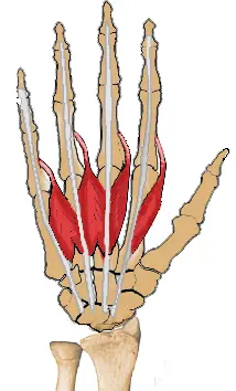
The lumbricals are intrinsic muscles of the hand that flex the metacarpophalangeal joints and extend the interphalangeal joints. They are four, small, worm-like muscles on each hand. These muscles are unusual in that they do not attach to bone.
This muscle appears to be rather weak based on its modest cross-sectional area and strength assessments from biomechanical investigations. This is particularly evident when contrasting the lumbrical with the interosseous muscle, which functions similarly but is much stronger.
The lumbricals’ high number of muscle spindles raises the possibility that these muscles are crucial for proprioceptive monitoring of the fingers. Additionally, the distribution of spindle fibres within the lumbricals and anatomical characteristics indicate that this muscle is more engaged in sensory feedback, which is critical for accurate object handling and pinch actions.
Origin:
- 1st lumbrical arises from the lateral side of the tendon of 2nd digiti.
- 2nd lumbrical arises from the lateral side of the tendon of 3rd digiti.
- 3rd lumbrical arises from the lateral side of the tendon of the 3rd and 4th digits.
- 4th lumbrical arises from the adjacent sides of the tendon of the 4th and 5th digits.
Insertion:
It inserts into the extensor expansion.
Nerve supply:
The first and second lumbricals are innervated by the median nerve and the third and fourth, by the deep branch of the ulnar nerve.
Blood supply:
Four separate sources supplies to these muscles, they are
- Superficial palmar arch
- Common palmar digital artery
- Deep palmar arch
- Dorsal digital artery.
Action:
It flexes the metacarpophalangeal joints and extends interphalangeal joints.
Function:
The fact that the lumbrical muscles of the hand originate from tendons and insert into the extensor expansions, instead of bony structures, makes both of their attachment points quite mobile.
That means the muscles are capable of two different actions. These are flexion at the metacarpophalangeal joints and extension in both the proximal (PIP) and distal interphalangeal joints (DIP). The reason for the opposite actions is that the tendons cross the MCP on the palmar side but distally insert at the dorsal side of the finger.
These combined movements play a role in the complex movement of the hand (e.g. for holding a pen), and contribute to the general dexterity of the hand.
Strengthening exercise:
Bend fingers down until all knuckles are bent. The tips of the fingers should touch the pads at the base of the fingers. Hold the stretch for a few seconds and repeat with the opposite hand. Complete the stretch five times daily.
Stretching exercise:
Keeping the knuckles straight, bend fingers forward forming an L shape with the fingers and the hand. Hold for a few seconds and repeat with the opposite hand. Complete the stretch five times.
Assessment
The patient is asked to flex the metacarpophalangeal joints while maintaining the interphalangeal joints extended, with the palm supine, so that physicians may test the patient’s lumbrical muscle strength.
The proximal phalanx of each of the fingers 2 through 5 is then subjected to a counterforce by the clinician along its palmar surface. To assess the extension of the interphalangeal joints while preserving flexion of the metacarpophalangeal joints, resistance can also be applied individually on the dorsal side of the middle and distal phalanges of digits 2–5.
Clinical importance:
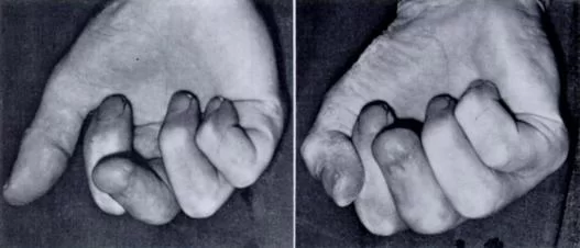
Lumbrical-plus finger
When the tendons of the flexor digitorum profundus detach distal to the origin points of the lumbricals an interesting phenomenon occurs: when trying to close the fist, the fingers strangely extend instead.
Crush Injury
Crush injuries involving the hand can occasionally cause injury to the lumbrical muscle. Adhesions between the interossous and lumbrical muscles may form after these injuries. Adhesions distal to the inter-palmar plate ligament restrict the proximal mobility of the interossous and lumbrical muscles because the lumbrical passes volar to the ligament while the interossous muscle passes dorsal.
When the patient makes a fist, the abnormality creates intermetcarpal discomfort. Usually, the affected muscles may be released to cure it. It has also been demonstrated that the lumbrical muscles have a role in carpal tunnel syndrome.
Etymology
Latin “lumbrical” means “worm” in its original language.
Variations
According to traditional instruction, the medial two lumbricals are innervated by the ulnar nerve and the lateral two lumbricals by the median nerve. Although about 50–60% of hands fit this description, 20–30% of hands only have median nerve innervation to the first lumbrical, and 20% of hands have median nerve innervation to the first three lumbricals.
Additionally, a cadaveric research discovered that in 64 percent of cases, the third lumbrical had dual innervation from both the median and ulnar nerves. The first lumbrical always receives its nerve supply from the median nerve, and the fourth lumbrical always receives its innervation from the deep branch of the ulnar nerve. However, innervation to individual lumbricals can vary greatly.
Two other physiological variants of the lumbrical muscles, discovered in around 10 percent of specimens in one research, are a bipennate second lumbrical and a double first lumbrical. Additionally, a third lumbrical was lacking in 2% of the cases in this investigation.
The lumbricals’ distal insertion locations exhibit a significant degree of diversity as well, with the variability increasing from the lateral to the medial lumbricals. These sites might be the extensor hood, the volar aspect of the metacarpophalangeal joint, the proximal phalanx, or any combination of these. The first lumbrical is more likely to have additional insertions into the extensor hood, whereas the fourth lumbrical is more likely to have attachments to the volar plate and bone.
FAQ
On each hand, the lumbricals are four tiny, worm-like muscles. The fact that these muscles are not attached to a bone makes them unique. Rather, they connect distally to the extensor expansions and proximally to the flexor digitorum profundus tendons.
Lumbricals are muscles in the hand, and they are associated with the second, third, and fourth fingers.
It has long been known that the ulnar nerve innervates the third and fourth lumbricals, whereas the median nerve innervates the first and second lumbricals.
The flexion at the corresponding metacarpophalangeal (MCP) joint and the extension at the proximal and distal interphalangeal (DIP) joints are among the lumbricals’ functions. It’s interesting to note that the flexor digitorum profundus, the lumbricals’ partial antagonist, is where they originate.
Hand
The lumbrical muscles are four short intrinsic hand muscles that are situated deep to the palmar fascia between the metacarpal bones. They acquire their name from their worm-like appearance (lumbricidae, Latin for earthworm).
References
Lumbricals of the hand. (2023, August 13). In Wikipedia. https://en.wikipedia.org/wiki/Lumbricals_of_the_hand