
Platysma muscle
Table of Contents
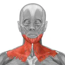
Platysma Muscle Anatomy
This muscle covers a portion of a neck muscle known as the sternocleidomastoid. The platysma muscle is expansive in size, with a broad width that spans the collarbone, or clavicle, and the side of the neck. Its point of origination is the upper portions of the pectoral, or chest, and the deltoid, or shoulder.
Origin
It originates from the upper parts of the pectoralis and deltoid fascia. Its fiber runs upwards and medially.
Insertion
It inserts into the anterior fibers to the base of mandible, posterior fibers to the skin of the lower face and lip and may be continuous with the risorius.
Nerve supply
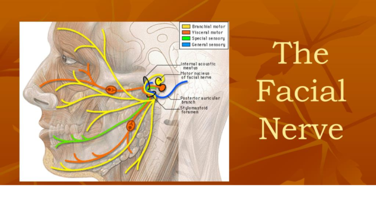
The platysma is supplied by the cervical branch of the facial nerve.
Blood supply
The platysma receives arterial blood supply from the submental branch of the facial artery as well as the suprascapular branch of the thyrocervical trunk.
Anatomical Variations
Up to one or two centimetres below the chin, 75% of the medial fibres in the submental region interdigitate with the contralateral platysma muscle. Ten percent of cases had no interdigitation of the medial platysmal fibres, whereas fifteen percent have interdigitation down to the thyroid cartilage.
Physiologic Variants
The origin and location of the platysma muscle fibres vary greatly. The muscle may not exist or may be quite thin in some people’s neck regions. In other situations, the fibres could terminate superiorly at the mental bone. Seldom can the platysma muscle fibres be observed to be as high as the zygomaticus or the orbicularis oris muscles’ borders.
Function
When the entire platysma is in action, it produces a slight wrinkling of the surface of the skin of the neck in an oblique direction.
Its anterior portion, the thickest part of the muscle, depresses the lower jaw; it also serves to draw down the lower lip and angle of the mouth in the expression of melancholy, i.e. surprise or horror.
However, the platysma plays only a minor role in depressing the lip which is primarily performed by the depressor anguli oris and the depressor labii inferioris.

It releases the pressure of skin on the subjacent veins depresses the mandible, and pulls the angle of the mouth downward as in horror or surprise.
Embryology
Beginning in weeks 9 and 10 of pregnancy, the cervical lamina and the superficial musculoaponeurotic system give rise to the platysma.
By week 17, the mandibular implantation site of the platysma is clearly visible. The parotid fascia is formed at this point when connective tissue and adipose tissue start to fill the area between the parotid’s capsule propia and platysma.
Platysma muscle Exercise
Strengthening Exercise:
1. Hanging-Head Exercise:
Lie on your back on a firm surface, such as a bed, with your head hanging slightly over the side. Rest your arms by your sides.
Exhaling, slowly lift your chin toward your chest.
Hold the contraction for a few seconds and then relax your head back to your starting position.
Repeat this exercise 10 times, increasing repetitions as your neck muscles grow stronger.
Chin-Firming Exercise:
Stand or sit with your shoulders relaxed, facing forward.
Place your lower lip over your upper lip and tilt your head back, feeling the stretch along the sides of the jaw and along the front of the neck.
Stretch until you can see the ceiling, and hold this position for several seconds.
Clinical Importance
In Bell’s palsy platysma muscle commonly paralysis.
Age-related changes to the platysma
As we age, the platysma loses definition and becomes thinner. When young, the muscle fibres frequently fuse with those on the opposing side. The medial fibres split as people age. The muscle weakens and sags, giving rise to the characteristic “platysmal bands.” Further laxity of the neck tissues is caused by ptosis of the muscle.
Relationship of SMAS to the platysma:
The platysma was traditionally shown as primarily a cervical muscle, with most anatomy textbooks only displaying a tiny extension of the platysma into the face beyond the mandibular line, with the SMAS being a separate facial component. According to recent anatomical and surgical studies, the platysma really spreads more superiorly into the face and contains a considerable facial component.
According to Mitz and Peyronie (1976), the SMAS is a fibro-fatty layer in the face with unique facial components. Studies have indicated that the platysma encompasses about 50% of the length between the mandibular angle and the malar eminence, sometimes referred to as the mandibular malar line, or MML. Because the SMAS and the platysma blend together so well, some have suggested that surgeons treat them as a composite flap rather than as separate structures in the face and neck, respectively.
In fact, when dealing with these tissues during cervicofacial rhytidectomy and neck lifts, it is probably wiser to conceive of the layer as the superficial musculoaponeurotic system-platysma complex (SMASP) rather than distinct parts.
Platysmal bands
Most people start to develop platysmal bands by the time they are 55 years old, and they get worse with time. The reason platysmal bands emerge with ageing is explained by a number of possibilities. Skin looseness, a decrease in platysmal tone, and the platysma’s separation from deeper attachments are a few of these. According to a more recent idea, skin laxity is not caused first by hyperkinetic platysma, but rather by it becoming more conspicuous in the shape of bands.
Since the anterior platysma bands deteriorate with time on the non-paralyzed side but are not visible in the presence of facial palsy, the current theory is that they are caused by the contracture of these anterior free platysmal fibres.
Trevidic et al.’s proposal for the treatment of platysmal bands involves selective denervation of the platysma, which is based on investigations they conducted on individuals who had unilateral facial paralysis.
Plastic surgery
Facial nerve palsy secondary consequence is wrinkled neck skin resulting from a loss in muscular tone that shortens and thins the muscle, a condition that can be linked to natural ageing. As one ages, neck bands in the region above the platysma muscle become more apparent. Weightlifting or facelift surgeries may make them worse. This might be referred to as “turkey neck” or platysma dyskinesia.
One could use conservative management. As alternatives, therapies include platysmaplasty and injections of botulinum toxin. This type of surgery is termed platysmaplasty, and it can be performed openly or closed. In the latter case, a plastymotome, a specialist tool, is utilized to perform the procedure without the need for incisions. The symptoms start to lessen after about two weeks.
Because adipose tissue is located above the platysma muscle, neck liposuction may be done without puncturing it. Preventing bleeding also requires avoiding injury to the platysma muscle.
FAQs
Neck
Unlike other muscles in the body that are situated close to the subcutaneous tissue, the platysma is found in the neck’s subcutaneous tissue, which is the cervical fascia’s superficial layer. Because of its superficial location, the underlying neurovascular systems must be acknowledged during surgical neck dissections.
At the front of the neck, the platysma muscle may be absent or interdigitate with the opposing side’s muscle; it is attached to the clavicle, mastoid process, or occipital bone.
The cervical branch of the facial nerve (38.2%) or the cervical branch and mandibular branch of the facial nerve (60.5%) innervated the platysma most frequently. The cervical plexus (0.6%), cervical motor nucleus (0.6%), and glossopharyngeal nerve (0.1%) were the next most common innervations.
To reduce the look of your double chin, platysma exercise can help tighten the muscles below and the loosen skin around it.
No, The platysma muscle performs a number of crucial tasks. These include widening the lips, tense neck muscles, and a range of emotions on the face, including surprise, grins, frowns, and fury.
At around 6 millimeters in length, the stapedius muscle is the smallest muscle in the human body. It connects the posterior portion of the stapes neck to the pyramidal eminence of the petrous region of the temporal bone and is situated in the tympanic chamber of the middle ear.
Exercises with platysmal bands have the potential to strengthen the neck muscles. This is said to lessen the possibility of platysmal banding.
References
- Hoerter, J. E. (2023, August 7). Anatomy, Head and Neck, Platysma. StatPearls – NCBI Bookshelf. https://www.ncbi.nlm.nih.gov/books/NBK545294/
- Platysma muscle. (2023, June 1). In Wikipedia. https://en.wikipedia.org/wiki/Platysma_muscle
4 Comments