
Rectus femoris Muscle
Table of Contents
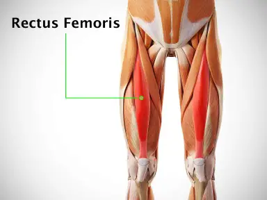
Rectus femoris Muscle Anatomy
The rectus femoris is a key muscle located in the anterior (front) compartment of the thigh. It is one of the four quadriceps muscles, along with the vastus lateralis, vastus medialis, and vastus intermedius.
As one of the primary extensors of the knee joint, the rectus femoris plays a crucial role in activities such as walking, running, and jumping. Additionally, it is involved in the flexion of the hip joint.
Unlike other quadriceps muscles, this one connects the hip and knee joints. It comes from the hip bone’s anterior inferior iliac spine and inserts into the patella and tibial tuberosity through the quadriceps tendon. The femoral artery supplies blood to the rectus femoris, which is innervated by the femoral nerve. Its importance in a variety of functional movements and activities is highlighted by its dual function in both hip flexion and knee extension.
Origin:
It originates in 2 heads. They are as follows:
Straight head: originates from the anterior inferior iliac spine
Reflected head: originates from a curved line along the upper part of the acetabulum at the ilium.
Insertion:
It inserts into the patellar tendon as one of the four quadriceps muscles.
Nerve supply:
The Posterior division of the femoral nerve (L3, L4) supplies the muscle.
The corticospinal tract is the pathway via which cortical signals from the precentral gyrus apex descend. In an area known as the “pyramidal tract decussation,” at the level of the lower medulla, over 90% of the fibers in this nerve tract decussate. Up until they get to the lower motor neurons beneath the upper thoracic spinal cord, these fibers continue to descend on the contralateral side as the lateral corticospinal tract. Although they do not innervate structures below the upper thoracic spinal cord, the remaining 10% of the fibers that do not decussate continue as the ventral corticospinal tract.
The fibers of the lateral corticospinal tract synapse with the lower motor neurons at the L4 level to reach the quadriceps femoris muscle. The neuronal signal then travels in the direction of the lumbar plexus and the L4 anterior rami.
L2 and L3 fibers contribute to the formation of the femoral nerve, which originates from the L4 anterior rami. All four of the quadriceps femoris’s components—including the rectus femoris—are innervated by the femoral nerve.
Blood supply:
The descending branch of the Lateral Circumflex Femoral Artery supplies to the muscle.
Lymphatics Drainage
The femoral vein and its branches carry blood out of the muscle.
The superficial inguinal nodes receive the drainage from the anterior thigh’s lymphatic veins. The arteries then climb to the lymph nodes in the lateral aortic and external iliac branches.
Action:
It acts as knee extensor and hip flexor.
Function of the Rectus Femoris
Along with the sartorius and iliopsoas, the rectus femoris flexes the hip and, as part of the quadriceps femoris, stretches the knee. At the knee and hip, this superficial muscle irritates the hamstrings.
Multi-joint muscles can develop restrictions in function, both passive and active. Active insufficiency in the rectus femoris happens when the knee’s capacity to fully extend restricts the hip’s range of motion, and vice versa. In this state, the muscle is strongly contracted. When the hip is extended while seated, the rectus femoris weakens as a knee extensor.
Conversely, when the rectus femoris’s ability to extend the hip and vice versa is limited by complete knee flexion, this is known as passive insufficiency of the rectus femoris. The muscle is at its longest in this instance.
Structure of the Rectus Femoris
With deep, vertically directed fibers and superficial, bipenniform fibers, the rectus femoris has a fusiform morphology. This muscle comes from two places. The anterior inferior iliac spine is the origin of the “direct head,” whereas the superior acetabular ridge is the origin of the “indirect head.” A common tendon is formed when the muscle heads join distally at an acute angle.
Through the quadriceps tendon, the rectus femoris inserts jointly with the other quadriceps muscles on the patellar base inferiorly. The tibial tuberosity serves as an additional point of insertion for the quadriceps muscle complex due to the relationship between the patella, patellar ligament, and quadriceps tendon.
Because it crosses two lower limb joints—the thigh and knee—and is superficially located, the rectus femoris can be easily identified from the other quadriceps muscles.
Relations
The sartorius, iliacus, and tensor fasciae latae muscles are situated deep to the proximal portion of the rectus femoris muscle. The anterior compartment of the thigh is completely filled up, all the way up to the rectus femoris. These consist of the lateral circumflex femoral artery, the vastus intermedius, the hip joint capsule, the anterior borders of the vastus lateralis, and a few femoral nerve branches.
Embryology
The fifth week of development is when the lower limb bud emerges. The ectoderm, endoderm, and mesoderm are the three basic germ layers that make up the bud, which emerges laterally from the L2 to S2 spinal segments. The myotomic sections of somites, which originate from the mesoderm, give rise to the rectus femoris muscle tissue.
In the meantime, the lateral plate somatic mesoderm produces the skeletal components that support the lower leg. The lower limb rotates 90 degrees medially about the longitudinal axis throughout development, bringing the knee to the fetus’s anterior aspect.
Rectus femoris Muscle Exercise
Strengthening Exercises:
1. Straight Leg Raise Exercise:
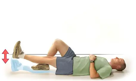
- The straight leg raising exercise is a simple way to get your quad muscles working properly.
- Lie on your back on a flat surface.
- Bend the knee of your unaffected leg to a 90-degree angle and keep your foot flat on the surface.
- Keep your other leg straight without the knee bent and point your toes toward the ceiling.
- Slowly lift the involved leg off the floor by contracting the front thigh muscles.
- Hold for five seconds.
- Slowly lower your leg to the floor. Relax, then repeat 10 to 15 times.
2. Wall Slide Squats:
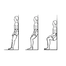
- Stand upright with your back against a wall and feet shoulder-width apart.
- Slowly bend your knees, sliding your back down the wall for a count of five until your knees are bent at a 45-degree angle. (Do not bend too much further than this, as it will cause increased strain on your knees.) Hold this position for five seconds.
- Straighten your knees by slowly sliding up the wall until you are fully upright with your knees straight.
- Repeat the above steps 10 more times.
Stretching Exercises:
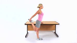
- This can be performed in either standing, or laying on your front.
- Pull the foot of the injured leg towards your buttock until you can feel a gentle stretch on the front of the thigh.
- To increase the stretch, tilt your hips backwards.
- Hold for 20-30 seconds and repeat 3 times.
- Do this at least 3 times a day.
Anatomical Variantions
The rectus femoris can give rise to accessory muscles. One of these variations is a muscle slip that inserts straight into the vastus lateralis from the acetabulum. The proximal origin of the rectus femoris muscle may alternatively have just one head rather than two, with the acetabular origin absent and the lone tendon originating from the lower anterior iliac spine.
Clinical Importance
The quadriceps is the most commonly injured muscle in sports that require sprinting or kicking, such as soccer. The rectus femoris (RF) is the most commonly injured quadriceps muscle especially in young athletic individuals.
Heart failure (HF) is often accompanied by skeletal muscle weakness and exercise intolerance, which are known as prognostic factors of HF. Comprehensive evaluation of physical function is important, but it is not commonly conducted because of the lack of equipment or appropriate expertise. Measurement of rectus femoris diameter (RFD) by ultrasound is convenient and noninvasive, but it has not been clarified that RFD could represent physical functions in HF patients.
Acute rectus femoris muscle injuries manifest as sudden, intense pain that tears easily. In contrast, subacute injuries appear gradually and typically start out as mild-to-moderate pain that gets worse when you run. Both MRIs and ultrasonography are useful for diagnosing and tracking damage to this muscle.
While they are rare, proximal rectus femoris rips are linked to sports like soccer and other activities where players must kick and sprint a lot. Compared to the indirect head, the direct head is more frequently involved. Both are present in total avulsions. Conservative care can be used to treat acute single-head proximal rectus femoris injuries, despite the fact that they frequently reoccur. Recurrent single-head rips and complete avulsion should be treated surgically. The prognosis for both disorders is favorable, with patients able to resume their pre-injury functional levels following surgery.
Avulsion fracture
An Avulsion fracture is the result of a portion of the anterior inferior ilac spine of the hip (AIIS) avulsing due to the Rectus femoris tendon. This results from a strong muscle contraction that pushes against the force holding the bone together. This is a common, yet uncommon, sports condition that can harm young athletes.
FAQ
The muscle closest to the skin and almost vertically positioned in the anterior thigh compartment is the rectus femoris. One of the most significant dynamic stabilizers of the knee is the quadriceps muscle complex, which includes this bipennate structure.
The strongest muscle in the human body is the quadriceps femoris, or quad muscle as it is popularly called. Along with the sartorius, it is housed in the anterior compartment of the thigh. The Latin term quadriceps femoris means “four-headed muscle.”
Most of the other three quadriceps muscles are covered by the rectus femoris muscle, which is located in the center of the thigh. It comes from the ilium. Its straight route gives it its name. On the lateral side of the femur, or the outside of the thigh, is the vastus lateralis muscle.
Being a biarticulate muscle, the rectus femoris crosses over the hip and knee joints. Although its primary use is as a knee extender, this muscle can also be used as a hip flexor due to its proximal attachments at the anterior inferior iliac spine and the acetabulum.
The only portion of the quadriceps muscle that attaches above the hip joint is the Rectus Femoris.
References
- Murdock, C. J. (2023, November 13). Anatomy, Abdomen and Pelvis, Rectus Femoris Muscle. StatPearls – NCBI Bookshelf. https://www.ncbi.nlm.nih.gov/books/NBK539897/
- Rectus femoris muscle. (2023, October 16). In Wikipedia. https://en.wikipedia.org/wiki/Rectus_femoris_muscle
3 Comments