Bones Of The Arm
Table of Contents
Introduction
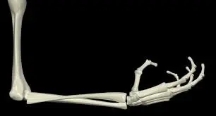
A human arm is composed of three bones, namely – the humerus, ulna, and radius. The human arm is an essential component that allows movement along the shoulder, elbow, wrist, and fingers, which is helpful for daily tasks.
The humerus, ulna, and radius are the three bones that make up an arm in humans. The human arm is an essential component that allows movement along the shoulder, elbow, wrist, and fingers, which is helpful for daily tasks.
The body’s upper limbs are the arms. These are among the most complex and often utilized bodily parts.
Each arm consists of four main parts:
- Upper arm
- Forearm
- Wrist
- Hand
Bones Of The Arm and Joints: Anatomy and Functionality
Bone of the Upper arm:
The space between the shoulder and elbow joint is referred to as the upper arm. The following are among the upper arm’s bones:
Scapula: Another name for the scapula is the shoulder blade. It is a flat, triangle-shaped bone that is primarily joined to the body by muscle. The arm and torso are joined by it.
Clavicle: Another name for the clavicle is the collarbone. It joins the arm to the body, just like the scapula does. It also facilitates the transfer of force from the arm’s upper region to the skeleton as a whole.
Humerus: The long bone of the upper arm is called the humerus. It is situated between the elbow joint and the scapula. The humerus is the attachment point for numerous arm muscles and ligaments.
- Additionally, the upper arm has a number of joints, such as the:
Acromioclavicular joint: This joint is where the clavicle and scapula meet.
Glenohumeral joint: The scapula and humerus converge at this joint.
Sternoclavicular joint: This joint is where the clavicle and sternum, or breastbone, attach.
Bone of the Forearm:
- The region between the elbow joint and the wrist is known as the forearm. The ulna and radius are its two main bones:
Radius: The side of the forearm nearest to the thumb is where the radius is found. Depending on how the hand is moved, it can twist around the ulna and change positions. The elbow, wrist, and finger joints can move more easily thanks to the numerous muscles that are linked to the radius.
Ulna: The radius and the ulna are parallel. The side of the forearm nearest the pinky finger is where it is located. The ulna does not twist and is immobile in contrast to the radius.
- The upper arm’s humerus bone joins the radius and ulna bones of the forearm at the elbow joint.
- In reality, the elbow joint is made up of three different joints:
Ulnohumeral joint: This is where the ulna and humerus join.
Radiocapitellar joint: The radius and the capitellum, a humerus region, merge at this joint.
Proximal radioulnar joint: The ulna and radius are joined at this joint, which permits hand rotation.
Bones Of The Arm Related Muscles
Upper arm:
- The anterior and posterior compartments are the two compartments that make up the upper arm.
Muscle movement:
- In advance of studying the various muscles, it’s critical to comprehend the four main motion patterns that each muscle performs:
Flexion: For example, this movement pushes the upper arm and forearm closer together.
Extension: The distance between two body parts is increased by this movement. The elbow being straight is one way to do this.
Abduction: Lifting an arm out and away from the body is an example of moving a body part away from the center of the body.
Adduction: It is the movement of a bodily part towards the center of the body, as in the case of the arm being brought back in to rest along the torso.
Anterior compartment:
- The primary bone of the upper arms, the humerus, is situated in front of the anterior compartment.
- The anterior compartment muscles consist of the following:
Biceps brachii: This muscle, which is sometimes called the biceps, has two heads that begin at the front and rear of the shoulder and unite at the elbow. The forearm is brought towards the upper arm by flexing the end that is closest to the elbow. The flexion and adduction of the upper arm are assisted by the two heads located near the shoulder.
Brachialis: This is the muscle that sits below the biceps. It serves as a link between the ulna, one of the forearm’s primary bones, and the humerus. It has to do with bending the forearm.
Coracobrachialis: This muscle can be found near the shoulder. It enables upper arm adduction and shoulder flexion. It also aids in the stabilization of the humerus within the shoulder joint.
Posterior compartment:
- The posterior compartment, which is positioned behind the humerus, is made up of two muscles:
Triceps brachii: This muscle, often known as the triceps, runs along the humerus and enables for forearm flexion and extension. It also helps keep the shoulder joint stable.
Anconeus: This is a tiny, triangular muscle that aids in elbow extension and forearm rotation. It’s sometimes thought to be a triceps extension.
Forearm:
- There are more muscles in the forearm than in the upper arm. It has two compartments, one anterior and one posterior, each of which has additional layers.
Anterior compartment:
The inside of the forearm is home to the anterior compartment. This region’s muscles are mostly used for forearm rotation and flexion of the wrist and fingers.
Superficial layer
Flexor carpi ulnaris: The wrist is flexed and adducted by this muscle.
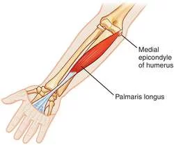
Palmaris longus: This muscle, which not everyone possesses, aids in wrist flexion.
Flexor carpi radialis: In addition to the abduction of the hand and wrist, this muscle permits the bending of the wrist.
Pronator teres: By rotating the forearm, this muscle permits the palm to face the body.
Intermediate layer
- Flexor digitorum superficialis: The second, third, fourth, and fifth fingers are flexed by this muscle.
Deep compartment:
Flexor digitorum profundus: This muscle aids in finger flexion as well. It also involves bringing the wrist closer to the torso.
Flexor pollicis longus: The thumb is flexed by this muscle.
Pronator quadratus: This muscle aids in forearm rotation, much like the pronator teres.
Posterior compartment:
The top of the forearm is where the posterior compartment is located. The wrist and fingers can be extended by the muscles in this compartment.
It contains an intermediary layer, in contrast to the anterior compartment.
Superficial layer
Brachioradialis: The forearm is bent at the elbow by this muscle.
Extensor carpi radialis longus: At the wrist joint, this muscle aids in the abduction and extension of the hand.
Extensor carpi radialis brevis: This muscle is the extensor carpi radialis longus’s shorter, broader equivalent.
Extensor digitorum: Extension of the second, third, fourth, and fifth fingers is possible due to this muscle.
Extensor carpi ulnari: The wrist is adducted by this muscle.
Deep layer
Supinator: The forearm can rotate outward such that the palm faces up thanks to this muscle.
Abductor pollicis longus: The thumb is abducted by this muscle, which pulls it away from the body.
Extensor pollicis brevis: The thumb is extended by this muscle.
Extensor pollicis longus: This is the extensor pollicis brevis’s lengthier equivalent.
Extensor indices: This muscle lengthens the index finger.
Bones Of The Arm Nerves
Brachial plexus:
- A collection of nerves that supply the skin and muscles of the arm is known as the brachial plexus. It travels down the arm after starting in the spine.
- There are five divisions within the brachial plexus:
Roots: This is where the brachial plexus starts. The C5, C6, C7, C8, and T1 spinal neurons combine to produce the five roots.
Trunks: The brachial plexus roots are made up of three trunks. The superior, middle, and inferior trunks are among them. The C5 and C6 roots combine to form the superior trunk, the C7 root continues in the middle trunk, and the C8 and T1 roots combine to form the inferior trunk.
Divisions: There are anterior and posterior divisions in each of the three trunks, for a total of six divisions.
Cords: Three cords—the lateral, posterior, and medial cords—are formed when the anterior and posterior divisions of the brachial plexus come together.
Branches: The peripheral nerves that supply the arm are formed by the brachial plexus branches.
Peripheral nerves:
- The arm’s peripheral nerves supply it with motor and sensory abilities.
- The following are among the six arm peripheral nerves:
Axillary nerve: Between the scapula and the humerus is where the axillary nerve passes. It activates the deltoid, teres minor, and partial triceps, among other shoulder-related muscles.
Musculocutaneous nerve: The biceps, brachialis, and coracobrachialis muscles are stimulated by this nerve, which passes in front of the humerus. The outside of the forearm is also sensed by the musculocutaneous nerve.
Ulnar nerve: On the outside of the forearm is where the ulnar nerve is situated. It gives the pinky and a portion of the ring fingers feeling as well as activating many hand muscles.
Radial nerve: The radial nerve passes through the inside of the forearm and behind the humerus. It activates the hand and wrist muscles as well as the triceps muscle in the upper arm. It gives the thumb’s proximal portion a feeling.
Median nerve: The median nerve runs the length of the arm’s interior. The majority of the hand, wrist, and forearm muscles are stimulated. Additionally, it gives feeling to a portion of the ring finger, middle finger, forefinger, and thumb.
Blood supply to Bones Of The Arm
- Numerous significant veins and arteries can be seen in each arm. Blood is transported from the heart to various parts of the body by arteries, while veins bring blood towards the heart.
- Some of the arm’s principal arteries and veins are listed below.
Upper arm and Forearm blood vessels:
Subclavian artery: The upper arm receives blood supply from the subclavian artery. It starts at the heart, passes beneath the collarbone, and ends up at the shoulder.
Axillary artery: The axillary artery receives its continuation from the subclavian artery. It is located beneath the underarm and provides blood to the shoulder region.
Brachial artery: The brachial artery receives its continuation from the axillary artery. It descends the upper arm and divides at the elbow joint into the radial and ulnar arteries.
Axillary vein: Blood from the shoulder and armpit region is transported to the heart by the axillary vein.
Cephalic and basilic veins: These veins pass through the upper arm and ascend. Eventually, they merge into the axillary vein.
Brachial veins: Large brachial veins that run parallel to the brachial artery can be found.
Radial artery: The forearm and hand receive blood supply from two arteries, one of which is this one. It moves along the forearm’s inside side.
Ulnar artery: The second of the two vessels that supply blood to the hand and forearm is the ulnar artery. It moves down the forearm’s outside.
Radial and ulnar veins: The radial and ulnar arteries are parallel to these veins. At the elbow joint, they merge with the brachial vein.
Common Arm Problems
The arms are two of the most used body parts, making them susceptible to a wide range of health issues. Here are a few examples of
the key ones.
Nerve injuries:
- There are several ways to harm the arm’s nerves, such as stretching, pinching, or cutting. These wounds may develop gradually over time or suddenly as a result of trauma.
- Although the location and type of the injury determine the precise symptoms, common signs of a nerve injury include:
- Pain that may be felt at the location of the damage or any place along the nerve
- Numbness or tingling in the arm or hand weakness in or around the injured area discomfort, which can be at the location of the injury or anywhere down the nerve
- Carpal tunnel syndrome and medial tunnel syndrome are two instances of arm nerve disorders.
Fractures:
- When a bone cracks or breaks as a result of trauma or injury, it is called a fracture. It is possible for any bone in the forearm or upper arm to fracture.
- The following are signs of an arm fracture:
- Pain or soreness in the arm
- Arm swelling
- Bruises where the injury occurred
- Restricted arm range of motion
Joint problems:
- Numerous issues can affect the joints of the upper arm and forearm, including the elbow and shoulder.
Joint problems can be brought on by inflammation, trauma, and repetitive use. - Among the common signs of an issue with the arm joint are:
- Discomfort in the affected joint
- Restricted mobility or stiffness in the impacted joint
- Swelling or inflammation in the damaged joint
- Among the conditions affecting the arm joints are bursitis, arthritis, and tennis elbow.
Vascular problems:
- Compared to the legs, the arms are less likely to experience vascular issues.
- When they do happen, several factors can contribute to their occurrence, such as atherosclerosis, plaque buildup on the artery walls, or blockages in the artery caused by blood clots.
- The following are signs of a vascular problem affecting the arm:
- Irritation, spasms, or pain in the afflicted arm
- A sensation of debility in the injured limb
- A feeling of being heavy in the afflicted arm
FAQs
Parts and Bones of the Pectoral Girdle
The humerus, or upper arm bone, and the ulna and radius, or forearm bones, make up your arm.
The ulnar nerve, which leaves the spine and passes through the neck, elbow, and fingers, is the nerve responsible for the so-called “funny bone.”
There are thirty bones in each arm of the human body.
The human arm consists of thirty bones. It has eight carpals (wrist bones), five metacarpals (palm bones), fourteen phalanges (five digits), one humerus (arm), one ulna, and one radius (forearm).
Arm
The triceps, which originates on the humerus and joins to the ulna at the elbow, is the muscle that extends or straightens the arm; the brachialis and biceps muscles work to bend the arm at the elbow. The ulna and radius are covered by several lesser muscles that move the hand and fingers in different ways.
Arm bones
The humerus, the upper arm bone, and the ulna and radius, the two forearm bones, make up your arm. Any of these bones can fracture, and the phrase “broken arm” can apply to any of them.
The humerus
The longest and largest bone in the upper extremity, extending from the shoulder to the elbow, is the humerus. It links the scapula and the lower arm, which is divided into three portions and is made up of the radius and ulna.
References:
1 Seladi-Schulman, J. 2018, August 27. Arm. Healthline. https://www.healthline.com/human-body-maps/arm.
2 Admin. 2022, August 25. Bones of the Arm – Diagram and Features BYJUS. https://byjus.com/biology/arm-bones-diagram/.