Distal Radius Fracture
Table of Contents
Introduction
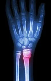
A distal radius fracture, or wrist fracture, is a break in the radius bone near the wrist. Pain, bruising, and sudden swelling are among the symptoms. The ulna bone may be broken as well.
The radius is one of two forearm bones that are found on the thumb side. The distal radius is the radius part that is attached to the wrist joint. When the radius breaks near the wrist, this is referred to as a distal radius fracture.
The radius’s distal end is defined as three centimeters proximal to the radiocarpal joint, where the radius meets the lunate and scaphoid bones of the wrist. A fracture is usually caused by falling on an outstretched arm. The vast majority of distal radial fractures are closed injuries with intact overlying skin. It can also occur in a car accident, a bike accident, a skiing accident, or another sporting event. The most common broken bone in the arm is the radius.
A fracture can be isolated, indicating that no other fractures are present. It can also occur in conjunction with a distal ulna fracture (the forearm bone on the small finger side). The injury is known as a distal radius and ulna fracture in these cases.
Classification of the distal radius fracture
The fracture is classified as a Colles or Smith fracture based on the angle of the distal radius as it breaks.
A Colles fracture can occur as a result of a direct impact on the palm, such as when you use your hands to break up a fall and land on your palms. The shape of a fork facing down is sometimes compared to the side view of a wrist after a Colles fracture. The wrist has a distinct “bump” similar to the fork’s neck. It occurs as a result of the broken end of the distal radius shifting up towards the back of the hand.
Smith fractures are the rarer of the two. It could be caused by a blow to the back of the wrist, such as falling on a bent wrist. In this type of fracture, the distal radius usually shifts down towards the palm side. This usually results in a noticeable drop in the wrist where the longer part of the radius terminates.
A Barton fracture can be volar (more common) or dorsal (less common), depending on whether the radius’s volar or dorsal rim is involved.
This is an intra-articular fracture of the distal radius with radio-carpal joint dislocation.
Causes of the distal radius fracture
A fall onto an outstretched arm is the most common cause of a distal radius fracture.
A relatively minor fall can result in a broken wrist due to osteoporosis (a common disorder in older adults in which bones become very fragile and more likely to break). A fall from a standing position causes many distal radius fractures in people over the age of 60.
A broken wrist can occur even in healthy bones if the trauma is severe enough. A car accident or a fall off a bike, for example, may generate enough force to break a young and healthy person’s wrist.
In older patients, good bone health may help to prevent fractures. People who have a family history of osteoporosis should consult their primary care physician about bone-strengthening options. More information: Calcium, Diet, and Bone Health.
Symptoms of the distal radius fracture
A broken wrist usually causes severe pain, tenderness, bruising, and swelling right away. In many cases, the wrist hangs in an unusual or bent position (deformity).
The nerve(s) to the hand can be injured in very severe fractures, resulting in numbness in the fingers. If you have numbness in your fingers after a wrist injury, go to an urgent care centre or emergency room as soon as possible. To avoid permanent nerve damage, the injury may need to be treated as soon as possible.
Diagnosis
If the injury is not too painful and the wrist is not deformed, you may be able to see a doctor the next day. An ice pack can be placed on the wrist, and it can be elevated until a doctor can examine it.
If the injury is extremely painful, the wrist is severely deformed, or the fingers are numb or pale, go to an urgent care centre or emergency room right away.
The doctor will most likely order wrist X-rays to confirm the diagnosis. X-rays can reveal whether or not a bone is broken and whether or not there is displacement (a gap between broken bones). They can also show how many broken bone pieces there are.
A computed tomography (CT) scan, which provides 3-D images of the broken bone, may be ordered in some cases by the doctor. This can be useful in surgical planning.
Physical examination
Physical therapists must perform a thorough physical exam that includes both subjective and objective data.
The subjective examination includes any information provided by the patient, such as pain, wrist ROM limitations, and activity limitations.
Wrist and digit ROM, grip, and forearm strength, bony and soft-tissue abnormalities, skin integrity, and nerve involvement are all evaluated objectively. Keep in mind that the contralateral extremity may be an untrustworthy control.
To avoid poor functional outcomes and prolonged recovery, healthcare professionals should evaluate ligamentous integrity as soon as possible in the presence of persistent pain associated with suspected carpal instability. Intercarpal ligament involvement is strongly indicated by specific fracture patterns and high-energy injuries.
Treatment of distal radius fracture
Treating broken bones follows a simple rule: the broken pieces must be repositioned and prevented from moving until they heal.
Many factors may affect how a distal radius fracture is treated, including:
- Displacement of fractures (whether or not the broken bones shifted)
- Comminution (the presence of fractures in multiple locations)
- Participation is required.
- Associated ulna fracture and median nerve injury
- Whichever hand is dominant
- Your occupation and level of activity
In any case, the immediate fracture treatment uses a splint to provide comfort and pain relief. If the fracture is displaced, it is reduced before being splinted. Fracture reduction is done under local anaesthesia, which numbs only the painful area.
There are multiple techniques for treating a distal radius fracture. Many factors influence the decision, including the nature of the fracture, your age and activity level, and the surgeon’s best judgment.
Non-surgical treatment
If the broken bone is in an ideal position, a cast may be specific until the bone heals. If your broken bone is out of place and likely to limit future use of your arm, it may be necessary to re-align the broken bone fragments. The process by which the doctor transfers the broken pieces into position is known as reduction.
A splint or cast may be placed on your arm after the bone has been properly aligned to keep the bones aligned. For the first few days, a splint is usually used to allow for some normal swelling. After the swelling has subsided, a cast is usually applied a few days to a week later. The cast is frequently changed after 2 or 3 weeks because the cast loosens as the swelling decreases.
Depending on the nature of the fracture, your doctor may take regular X-rays to closely monitor the healing process. X-rays are frequently taken at weekly intervals for 3 weeks and then again at 6 weeks in patients treated without surgery. If the fracture did not need to be reduced and/or was thought to be stable, X-rays may be taken less frequently. If the fracture becomes misaligned at any point, surgery may be recommended.
The cast is typically removed 6 weeks after the fracture in non-operative fractures. You will most likely begin physical therapy at that point to help improve the motion and function of the injured wrist. To protect the healing bone, you will typically wear a removable splint between therapy sessions.
Surgical treatment
Sometimes the bone position cannot be corrected with a closed reduction alone and/or cannot be maintained in a cast. These fractures may heal out of alignment, resulting in poor arm function. In such cases, surgery may be required to correct the fracture and stabilize it while it heals.
Procedure
To access the broken bone(s), an incision on the wrist over the fracture is typically made. Important structures like arteries, nerves, and tendons are identified and safeguarded. The broken bone(s) are realigned directly through the incision by the surgeon. This is also referred to as an open reduction.
There are several options for holding the bone in the correct position while it heals after it has been re-aligned, depending on the fracture:
- Casts are rarely used following an open reduction.
- Metal pins (usually stainless steel or titanium) — This treatment was popular many years ago and is still commonly used in children with growing bones.
- Plate and screws — The most common treatment option for distal radius fractures.
- External fixator (a stabilizing frame outside the body that holds the bones in place so they can heal) — This has become less common in recent years, but it is still a viable option for severe and open fractures.
- Any combination of these approaches
Open fractures.
Most open fractures require surgery as soon as possible (typically within 24 hours or less of injury).
To prevent infection, the exposed soft tissue and bone are thoroughly cleaned (debrided) and antibiotics are administered.
External or internal fixation methods are frequently used to keep the bones in place.
Your doctor may use a temporary external fixator if the soft tissues around the fracture are severely damaged.
Internal fixation with plates or screws may be used in a subsequent procedure after the swelling has subsided and the soft tissues around the fracture have healed.
Rehabilitation
After a distal radius fracture, most people resume all of their previous activities. The type of injury, the type of treatment received, and the body’s reaction to the treatment all have an impact. In some cases, such injuries can result in permanent functional limitations.
Almost all patients will experience wrist stiffness. This usually improves in a month or two of having the cast removed or having surgery, and it goes on getting better for a minimum of two years. If your doctor believes it is necessary, you will begin physical therapy within a few days to weeks of surgery, or immediately after the last cast is removed.
Most patients will be able to resume light activities, such as swimming or lower-body exercise in the gym, one to two months after the cast is removed or one to two months after surgery. Sports like skiing or football can be resumed between 3 and 6 months after the injury.
Complications
These are the most common complications associated with distal radius fractures:
Malunion can be treated with corrective osteotomy of the malunion, which occurs when poor realignment results in a shortened radius compared to the ulnar, resulting in reduced wrist motion, wrist pain, and reduced forearm rotation.
Compression of the median nerve, which is more common in patients who heal with a significant degree of malunion
Osteoarthritis, particularly when there is intra-articular involvement from the initial fracture
Summary
A distal radius fracture is a break in the radius bone near the wrist, causing pain, bruising, and swelling. It is often caused by falling on an outstretched arm or a fall from a standing position. The most common broken bone in the arm is the radius. The fracture can be isolated or in conjunction with a distal ulna fracture.
It is classified as a Colles or Smith fracture based on the angle of the distal radius as it breaks. A fall onto an outstretched arm is the most frequent reason.
The symptoms of a distal radius fracture include severe pain, tenderness, bruising, and swelling, and may result in numbness in the fingers. Diagnosis involves a thorough physical examination, including subjective and objective data.
Distal radius fractures are broken bones that need to be repositioned and prevented from moving until they heal.
Treatment depends on factors such as displacement, concentration, participation, ulna fracture, dominant hand, and occupation.
In non-surgical treatment, a cast may be used to keep the fracture in place, while surgery may be necessary for open fractures. The procedure involves an incision on the wrist, realigning the broken bones through an incision. Various options for holding the bone in the correct position include casts, metal pins, plates, screws, or external fixators.
After surgery, most patients resume their previous activities, with wrist stiffness improving within a month or two. Common complications include malunion, compression of the median nerve, and osteoarthritis. Rehabilitation typically involves physical therapy and resuming light activities within a few months.
FAQs
Median nerve dysfunction caused by distal radius fractures can present acutely, subacutely, or late, and it can be caused by either primary or secondary causes.
The distal radius fracture is one of the most common wrist fractures. It usually happens when someone falls on their outstretched hand. A broken wrist is a break or cracks in one or more of your wrist’s bones.
The most common complication that occurs after a distal radius fracture is a malunion.
As a result, the most common type of “broken wrist” is a distal radius fracture (the larger forearm bone is broken near the wrist). This type of fracture is quite common. The radius is, in fact, the most commonly broken bone in the arm.
References:
- Distal Radius Fractures (Broken Wrist) – OrthoInfo – AAOS. (n.d.). https://orthoinfo.aaos.org/en/diseases–conditions/distal-radius-fractures-broken-wrist/
- Distal Radius Fracture (Wrist Fracture). (2021, August 8). Johns Hopkins Medicine. https://www.hopkinsmedicine.org/health/conditions-and-diseases/distal-radius-fracture-wrist-fracture
- Distal Radius Fractures – Colles – Smiths – Barton – TeachMeSurgery. (2021, October 15). TeachMeSurgery. https://teachmesurgery.com/orthopaedic/wrist-and-hand/distal-radius-fracture/
- Distal radius fracture. (2023, November 8). Wikipedia. https://en.wikipedia.org/wiki/Distal_radius_fracture
- Distal Radial Fractures. (n.d.). Physiopedia. https://www.physio-pedia.com/Distal_Radial_Fractures