Front of the forearm muscles
Table of Contents
Introduction
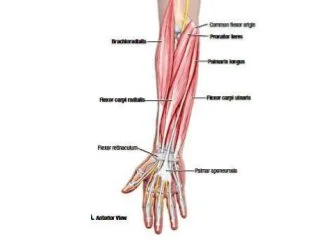
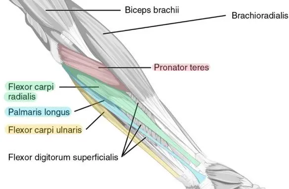
Superficial muscle of anterior compartment of the forearm
The muscles of the front of the forearm may be divided into superficial and deep groups of muscle.
There are five muscles in the superficial muscle group. These are the pronator teres, the flexor carpi radialis, the palmaris longus, the flexor carpi ulnaris and the flexor digitorum superficialis muscles.
Deep muscle of the anterior compartment of the forearm
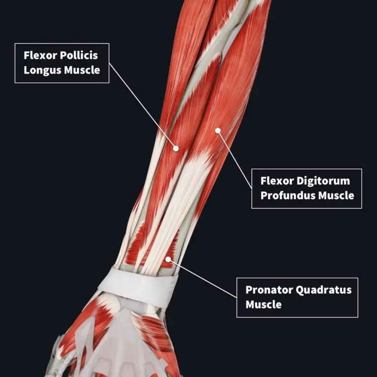
Deep muscles of the front of the forearm are the flexor digitorum profundus, the flexor pollicis longus, and the pronator quadratus muscles.
The forearm extends between the elbow joint and the wrist joint. Radius and ulna from its skeleton. Radius and ulna bones articulate at both their ends to form superior radioulnar joints and inferior radioulnar joints. Their shafts are kept at an optimal distance through the interosseous membrane.
Muscles accompanied by nerves and blood vessels are present on both sides on the front and the back of the forearm.
Superficial muscles of the front of the forearm
Pronator teres muscle
Origin
Humeral head: Humeral head of the pronator teres originate from the Medial epicondyle of the humerus
Ulnar head: Ulnar head of the pronator teres originate from the medial margin of the coronoid process of the ulna.
insertion
Pronator teres inserted on the Middle one-third of the lateral aspect of the shaft of radius
Nerve supply
The nerve supply of the Pronator teres is the Median nerve
Blood supply
The blood supply of the Pronator teres is the Branches of brachial, radial, and ulnar arteries
Action
It is the main pronator of the forearm. It also flexes the elbow.
Flexor carpi radialis
Origin
The origin of the Flexor carpi radialis is the Medial epicondyle of the humerus
insertion
Flexor carpi radialis muscle inserted into the palmar surface of the Bases of second and third metacarpal bones
Nerve supply
The nerve supply of the Flexor carpi radialis muscle is the Median nerve
Blood supply
The blood supply of the flexor carpi radialis muscle is an anterior ulnar recurrent artery or posterior ulnar recurrent artery
Action
Flexor of the wrist
Abductor of the wrist
Palmaris longus
Origin
The origin of the Palmaris longus is the Medial epicondyle of the humerus
insertion
The Palmaris longus is inserted on the Distal half of the Flexor retinaculum and the apex of the palmar aponeurosis
Nerve supply
The nerve supply of the Palmaris longus is the Median nerve
Blood supply
The blood supply of the Palmaris longus is Anterior ulnar recurrent artery, median artery
Action
Flexor of the wrist
Flexor carpi ulnaris
Origin
Humeral head
The humeral head of the Flexor carpi ulnaris muscle originates from the Medial epicondyle of the humerus
Ulnar head
The ulnar head of the Flexor carpi ulnaris muscle originates from the Medial margin of the olecranon, and from the posterior border of the ulna. The ulnar nerve passes between the humeral head and ulnar heads.
insertion
The Flexor carpi ulnaris muscle is inserted on the Pisiform bone, but the pull of the muscle is transmitted through the pisohammate and the base of the fifth metacarpal bone
Nerve supply
The nerve supply of the flexor carpi ulnaris muscle is the Ulnar nerve
Blood supply
The blood supply of the flexor carpi ulnaris muscle is the Posterior ulnar recurrent artery, ulnar artery
Action
Flexor of the wrist
Adduction of the wrist
Fixes the pisiform bone during contraction of the hypothenar muscles.
Flexor digitorum superficialis
Origin
Humeroulnar head
The medial epicondyle of the humerus, the ulnar collateral ligament, and a tubercle on the medial border of the coronoid process(ulna)
Radial head
The anterior border of the radius up to the insertion of the pronator teres muscle.
Some fibres arise from a fibrous arch passing from the ulna to the radius and connecting the two heads. the median nerve and the ulnar artery pass deep into this arch.
insertion
The Flexor digitorum superficialis muscle ends in four tendons, one each for the medial four fingers. Opposite the proximal phalanx, the tendon for each digit splits into medial and lateral slips which are inserted on the corresponding sides of the middle phalanx. At the wrist the four tendons are arranged in two pairs, the superficial pair for the middle and ring fingers, and the deep pair for the index and little fingers. The tendons lie medial to the palmaris longus muscle and lateral to the ulnar vessels and nerve.
The tendons enter the hand by passing deep to the flexor retinaculum, enclosed within a common synovial sheath(ulnar bursa).
Nerve supply
The nerve supply of the Flexor digitorum superficialis muscle is the Median nerve
Blood supply
The blood supply to the flexor digitorum superficialis muscle is the ulnar artery.
Action
Flexor of the proximal interphalangeal joints. It may also flex the metacarpophalangeal joints and wrist joints.
Deep muscles of the front of the forearm
Flexor digitorum profundus
Origin
Upper three-fourths of the anterior and medial surface of the ulnar shaft
Upper three-fourths of the posterior border of the ulna by aponeurosis.
Medial surface of the olecranon processes and coronoid processes of the ulna
Adjoin part of the anterior surface of the interosseous membrane
insertion
The flexor digitorum profundus muscle forms 4 tendons for the medial 4 digits which enter the palm by passing deep to the flexor retinaculum
Opposite the proximal phalanx of the corresponding digit, the tendon perforates the tendon of the flexor digitorum superficialis muscle
Each tendon is inserted on the palmar surface of the base of the distal phalanx is inserted on the palmar surface of the base of the distal phalanx
Nerve supply
Medial half by the ulnar nerve
Lateral half by the anterior interosseous nerve (C8, T1)
Blood supply
The blood supply of the flexor digitorum profundus is the anterior interosseous artery, which is a branch of the common interosseous artery and is accompanied by the palmar interosseous branch of the median nerve.
Action
Flexor of distal phalanges after the flexor digitorum superficialis muscle has flexed the middle phalanges
Secondarily, it flexes the other joints of the digits, fingers, and the wrist joint
It is the chief gripping muscle. It acts best when the wrist is in extension.
Flexor pollicis longus
Origin
Upper three-fourths of the anterior surface of the radius shaft.
Adjoin part of the anterior surface of the interosseous membrane.
Occasionally from the medial or lateral(or both)borders of the coronoid process of ulna
insertion
The Flexor pollicis longus tendon enters the palm by passing deep into the flexor retinaculum.
It is inserted into the palmar surface of the distal phalanx of the thumb.
Nerve supply
The nerve supply of the Flexor pollicis longus is the Anterior interosseous nerve
Blood supply
The Blood supply of the Flexor pollicis longus is the branch (anterior interosseous artery) of the ulnar artery.
Action
Flexes the distal phalanx of the thumb. Continued action can also flex the proximal joints crossed by the tendon.
Pronator quadratus
Origin
The Pronator quadratus originates from the Oblique ridge on the lower one-fourth of the anterior surface of the shaft of the ulna, and the area medial to it
insertion
The Pronator quadratus is inserted on the Superficial fibres into the lower one-fourth of the anterior surface and the anterior border of the radius, and Deep fibres into the triangular area above the ulnar notch
Nerve supply
The nerve supply of the Pronator quadratus is the Anterior interosseous nerve
Blood supply
The blood supply of the Pronator quadratus is the anterior interosseous artery
Action
Superficial fibres pronate the forearm
Deep fibres bind the lower ends of the radius and ulna
Clinical note
A chronic false tear of the superficial anterior muscles of the forearm often leads to inflammation and increased connective tissue in the common flexor tendon at the medial epicondyle of the humerus (medial epicondylitis).
Golfer’s elbow:
Golfer’s elbow (medial epicondylitis or pitcher’s elbow) is a tendinopathy caused by overuse or overload of the muscles of the forearm and affects the medial common flexor tendon of the elbow.
Common symptoms are pain that increases during wrist and hand movements and difficulty performing day-to-day tasks.
Physiotherapy treatment
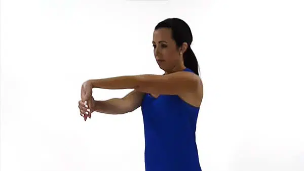
Stretching exercise
First Stretch your arm out in front of your body.
Slowly, point your fingers down until you can feel a stretch. Use the other hand to gently pull the raised hand toward your body. Hold this stretching position for 30 seconds and then relax.
Repeat this exercise three times.
Prayer position
Sit down with your palms together and your elbows on the table in a prayer position.
Lower the sides of the hands toward the table until you can feel a stretch. Keep your palms together. Hold this position for 30 seconds then Relax.
Repeat this exercise three times.

Strengthening exercise
Wrist curl
Sit down comfortably with your arm resting over your knees. Hold a weight with your palms facing up and your wrist hanging over the knee.
Move your hand up as far as you can and then down as far as you can in a slow and controlled motion.
Do 10 repetitions of 3 sets.

Strengthening exercise with a resistance band
Sit comfortably, resting your arm on a table with your palm facing up and your hand hanging over the edge of the table.
one end of the resistance band is put under your foot to hold it down and hold the other end in your hand. You may have to wrap it around your hand to produce some tension.
Pull up against the resistance, and flex your wrist as far as you can. Keep the motion smooth and controlled motion.
Slowly come back down to starting position.
Repeat 10 times.