Gluteus Medius Muscle
Table of Contents
Gluteus medeius Muscle Anatomy
Gluteus medius muscle is part of the three gluteal muscles and is a broad, thick, radiating muscle, situated on the outer surface of the pelvis.
The gluteal region, also referred to as the buttock is located posterior to the bony pelvis. The iliac crest defines its superior border, while the gluteal fold marks its inferior boundary. The quadratus femoris, piriformis, obturator internus, superior and inferior gemellus, and gluteus maximus, medius, and minimus are the muscles in that matter.
Between the gluteus maximus and gluteus minimus is the gluteus medius. The gluteus maximus covers the posterior third of it, while a robust layer of deep fascia simply covers the superficial anterior two-thirds.
Origin:
Outer surface of the ilium, between the anterior and inferior gluteal lines, and the edge of the greater sciatic notch.
Insertion:
Anterior surface of the greater trochanter of the femur.
Nerve supply:
Superior gluteal nerve L4, L5, S1).
The superior gluteal nerve supplies the gluteus medius. The dorsal branches of the sacral plexus nerve roots of the L4, L5, and S1 nerves give rise to the superior gluteal nerve. Via the larger sciatic foramen above the piriformis muscle, the nerve leaves the pelvis and divides into inferior and superior branches.
To innervate the gluteus minimus and gluteus medius, the superior branch of the deep division of the superior gluteal artery follows the upper branch. The inferior branch innervates the gluteus minimus and medius, finishing in the tensor fascia lata muscle, along with the lower ramus of the superior gluteal artery’s deep division.
Blood supply
The deep branch of the superior gluteal artery. The tendon is mainly supplied by the trochanteric anastomosis. This anastomosis is formed between the ascending branch of the medial circumflex femoral artery and the descending branches of the superior gluteal and inferior gluteal arteries.
Lymphatic Drainage
Blood is evacuated from the gluteal region by the superior gluteal vein (SGV), which has two branches: a superficial branch and a deep branch. The internal iliac vein is where the superior gluteal vein finishes after running parallel to the upper gluteal artery and entering the pelvis through the large ischial foramen in the supra-pyriform canal. There is an anastomosis between the superior and inferior gluteal veins.
The posterosuperior portion of the thigh and buttock is drained by the inferior gluteal vein (IGV). It comes from a single stem that runs parallel to the lower gluteal artery, where two branches converge. The inferior gluteal vein is anastomose with the medial circumflex veins of the femur and the superior perforating vein. The inferior gluteal vein drains into the distal section of the internal iliac vein after entering the pelvis through the lower opening of the big ischial foramen.
Action
Abduction of the thigh, internal rotation of the thigh.
Function
When the proximal attachment of the gluteus medius is fixed, the muscle can contract as a whole or it can contract with its anterior fibers only. In the former case, the axis of the movement goes through the hip joint and muscle pulls the greater trochanter superiorly and abducts the thigh.
In the latter case, as the axis of the movement tilts anteriorly, the muscle causes the internal rotation of the thigh.
Relations
Located superficially to the gluteus minimus and deep to the gluteus maximus, the gluteus medius is a middle gluteal muscle. The gluteus maximus covers only the posterior third of the muscle; the deep fascia of the hip covers the bigger anterior section of the muscle. There are instances where this fascia is called the gluteal aponeurosis. The piriformis muscle is situated anterior to the posterior margin of the muscle, and on occasion, the two may blend together.
The superior gluteal artery and nerve branches that pass between the gluteus medius and minimus muscles’ neighboring surfaces are connected to the gluteus medius. This relationship is significant because surgical treatments involving the incision of the gluteus medius may cause injury to these arteries and nerves.
Embryology
At approximately four weeks, or embryonic stage 14, the lower limb bud forms. At stage 17, the footplate of the lower limb has flattened, revealing a discernible hip joint but lacking a proper knee. The digits split from stages 20 to 23, with stage 23, or the end of week eight, marking the clear definition of the toes.
Cells produced from somites present at the level of the lower limb bud form the gluteus medius, just like other skeletal muscles. They then move into the bud of the limb and multiply there. They ultimately give rise to the gluteus medius muscle through the expression of myogenic determining factors.
Gluteus medius muscle exercise
Gluteus medius muscle exercise mainly of 2 types
- Gluteus medius muscle stretching exercise
- Gluteus medius muscle strengthening exercise
Strengthening exercise:
Clamshell exercise
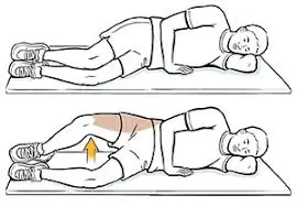
Lie on your side, with legs stacked and knees bent at a 45-degree angle.
Rest your head on your lower arm, and use your top arm to steady your frame. …
Engage your abdominals by pulling your belly button in, as this will help to stabilize your spine and pelvis.
Side lying hip abduction exercise
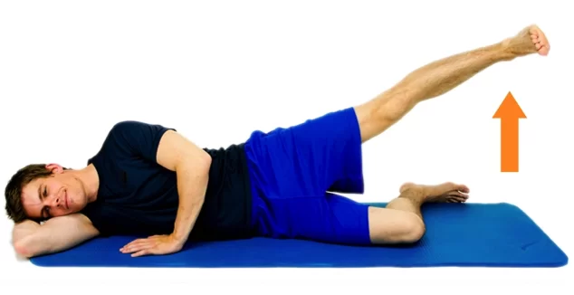
- Lie down on the floor on your side. Rest your head on your arm. Bend your legs at the knees.
- Keep your feet together and lift your top leg up so that your knees are separated. Keep your hips steady.
- Slowly lower your leg back down.
- Repeat 10 times, or as instructed.
- Switch sides if instructed.
1 Leg bridge exercise
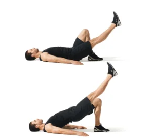
- Tighten your abdominal and buttock muscles.
- Raise your hips up to create a straight line from your knees to your shoulders.
- Squeeze your core and try to pull your belly button back toward your spine.
- Slowly raise and extend one leg while keeping your pelvis raised and level.
- Hold.
- Return to the starting position with knees bent.
- Perform the lift with the other leg.
Stretching exercise
Z – Sit exercise
- Begin by sitting comfortably on the ground.
- Bring your left knee to a 90-degree position in front of your body (as much as your body allows).
- Do the same with your right leg, toward the back of your body.
- You can sit upright in this pose or lean your torso forward toward your front leg.
- Hold the pose for 30 seconds, and then repeat on the other side.
Standing side bend exercise

- Using a wall for balance, stand with one side of your body on a wall.
- Cross your leg farthest from the wall in front of the other.
- Place one hand on the wall and the other on your hip. Then lean your upper body away from the wall, and push your hip toward the wall.
- Hold for 20 to 30 seconds, then repeat on the other side
Related pathology
A peripheral injury of the superior gluteal nerve may lead to loss of motor function. The classical sign is the pelvis dropping to the healthy side when standing on one leg (Trendelenburg’s sign). In order to maintain the balance, the patients compensatorily bend their upper body to the side of the stance leg. Furthermore, they walk with conspicuous sideward movements (Duchenne gait, also “waddling gait”).
Gluteus Medius Tears
When performing an intramuscular injection in the gluteal region, an injury of the sciatic nerve and superior gluteal nerve has to be avoided. Therefore, a recommended site of injection is the gluteus medius muscle in the upper outer quadrant of the buttock (Hochstetter’s technique).
Internal rotation of the thigh against resistance while in a supine posture with the hip and knee flexed is one way to assess the gluteus medius and minimus combined in a clinical context. An internal counter-resistance rotation test can be used to specifically detect the presence of gluteus medius tears. Clinical testing of the gluteus medius and minimus, as well as the tensor fascia lata, can be performed by having the patient abduct their leg against resistance while lying supine with their knee extended. Together, the gluteus medius and minimus support the pelvis.
Trendelenburg Gait
Patients with the gluteus medius and minimus paralysis will exhibit a distinctive Trendelenburg gait, which is a lurching gait. Running and walking will not be significantly affected by the paralysis of other muscles functioning on the hip joint if these two muscles and their innervations remain intact. Tendinopathy of the gluteus medius and/or minimus, with or without concurrent bursal disease, is the cause of greater trochanteric pain syndrome (GTPS). Often, patients report higher trochanter-localized lateral hip pain, which gets worse when they sleep on their sides or perform weight-bearing activities.
Clinically, the disease is identified by lateral hip pain and point tenderness at the greater trochanteric area. Walking, running, standing on one leg, and ascending stairs are examples of prolonged repeated activities that use the gluteus medius that causes pain. During an intramuscular injection in the gluteal region, harm to the superior gluteal nerve may occur. The sciatic nerve, which is often located in the lower quadrants of the gluteal region, and the superior gluteal should not be injured when injections are given in the upper lateral quadrant.
Gluteus medius syndrome (GMS)
There have been case reports of compression syndrome after trauma to the gluteus medius muscle, compressing the local vascular system, due to the close proximity of the musculature and developing neurovascular systems in the gluteal region.
Gluteus medius syndrome (GMS) shares similarities to greater trochanteric pain syndrome, however, it is a less prevalent source of leg pain or lower back pain. GMS can be correlated with failed back surgery syndrome and is linked to osteoarthritis at the hip or knee level as well as deterioration of the lumbar vertebrae.
Surgical Importance
Hip fractures, hip dislocations, and hip replacements all cause harm to the gluteus medius, as well as to the artery and nerve that supply it. The gluteus medius is surgically separated to provide access to the hip joint during a direct lateral approach to hip arthroplasty; hence, this procedure carries the highest risk of nerve injury. Even with soft tissue protecting cannulas, the deep superior branch of the superior gluteal nerve and its vessels are seriously endangered during the percutaneous placement of iliosacral screws, a common procedure used to treat complex pelvic injuries, even when the screws are positioned correctly.
Surgery may be necessary to treat gluteus medius tears, which can manifest as persistent trochanteric bursitis that is resistant to medication. The first step in diagnosing this pathology is usually a clinical evaluation, although MR imaging can also be helpful in this regard since it can show a high-intensity T2 signal that is related to the inflammation in the affected area. In this context, an additional useful diagnostic method for gluteal tendinopathy assessment is ultrasound.
Surgery can be performed endoscopically, utilizing a trans-tendon technique to optimize visualization and repair of the gluteus medius tear. If necessary, motorized burr can be used to remove the greater trochanter peak. It has also been suggested to treat gluteus medius tendinopathy with percutaneous ultrasonic tenotomy (PUT), a minimally invasive surgery that is frequently used to treat tendinopathies of the elbow, knee, and ankle. The method was tested on 29 patients by Baker et al., who showed improved hip clinical evaluation scores and good patient satisfaction.

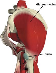
Gluteus medius muscle is the key muscle of Hip you should focus for regular strengthening and stretching exercise, This helps you walk, Regular Body weight transfer and also important for sports men who activity participate in High intensity activity.