
Gracilis Muscle
Table of Contents
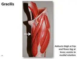
Gracilis Muscle Anatomy
The gracilis muscle is a long, thin muscle found in the thigh’s medial (adductor) compartment. Along with the adductor longus, adductor brevis, adductor magnus, and pectineus muscles, it is a member of the adductor muscle group.
The most superficial hip adductor, the gracilis, lies on top of the other four. Although it is the weakest component, the only hip adductor that crosses and works on the knee and hip joints is this one.
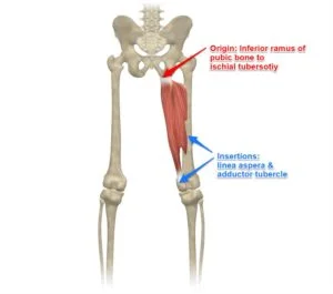
Origin of Gracilis muscle
It originates from the inferior rami of the pubis, and the body of the pubis.
Insertion
it attaches to the medial surface of the tibia, between the tendons of the sartorius and the semitendinosus.
Nerve supply
Gracilis muscle is innervated by the obturator nerve, which is a branch of the lumbar plexus. It arises from the L2-L3 spinal nerves.
Blood supply
Gracilis receives the majority of its vascular supply from the ‘artery to the adductors’, which is a branch of the deep femoral artery. The ‘artery to the adductors’ enters gracilis via its lateral surface, approximately one-third away from its origin.
Action of Gracilis muscle
flexion of the hip and knee joint, internal rotation of the knee, and adduction of the thigh.
Structure and Function
A long, thin muscle in the thigh’s medial compartment is called the gracilis. It comes from the ischiopubic ramus’ medial side. It is also connected to the inferior pubic ligament, which is a band that joins the inferior portions of the pubic rami, along with the adductor brevis muscle. The pes anserinus muscle tendon, which inserts on the superior medial tibia, medial to the tibial tuberosity, is formed distally by the gracilis muscle tendon joining forces with the sartorius and semitendinosus tendons.
The gracilis muscle is a spiral unipennate muscle because of the way its muscle fiber bundles are arranged. They obliquely insert into the anterior portion of the gracilis tendon.
The gracilis muscle has five to seven muscle fiber bundle compartments. Nerve branches run through each compartment, suggesting that each compartment may have a separate neuromuscular compartment function.
The gracilis muscle crosses the knee and hip joints and is responsible for internal rotation, knee flexion, hip adduction, and hip flexion.
Relations
The superficial muscle of the thigh’s medial (adductor) compartment is called the gracilis. The skin and subcutaneous tissue cover its superficial layer, and the deep layer of fascia lata covers its medial portion as well. The saphenous nerve emerges from the adductor canal and becomes subcutaneous, piercing the fascia lata between the gracilis and sartorius tendons. Additionally, the descending genicular artery’s saphenous branch runs between the sartorius and gracilis.
The muscles known as adductor magnus and adductor brevis are situated deep within the gracilis. Nonetheless, this muscle’s spiral trajectory may lead to a number of intricate and shifting relationships. As a result, the gracilis is situated medial to the adductor longus, adjacent to the lower border of the adductor brevis, and the medial border of the adductor magnus. The anserine bursa divides the tibial collateral ligament from the gracilis tendon, which is situated deep within it.
Strengthening exercise of Gracilis muscle
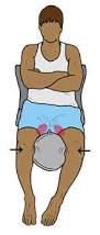
Sit on the floor with your feet together, hands on your feet, and elbows resting on your inner thighs. Gently press down on your thighs with your elbows until you feel a stretch through the inner thighs. Hold for 5 seconds. Now press up against the elbows with your thighs and hold for 5 seconds. Repeat the process back and forth for 30 seconds. Complete 3 sets of 30 seconds.
The butterfly groin stretch

Sit in an upright position. Place the soles of your feet together by bending your knees and rotating your thighs outward. A gentle should be felt in your groin and inner thigh as your knees lower down towards the floor. Hold the stretched position for 15 to 30 seconds, and then allow your knees to rise up, releasing the stretch. Repeat five times.
The groin squeezes for groin strength. Lie on your back with both knees bent. Place a rolled-up towel or pillow between your knees. Gently squeeze the towel with your inner knees, and hold this squeeze for five seconds. Slowly release. Repeat the exercise 10 times
Stretching exercise of Gracilis muscle
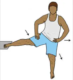
Place your right leg on a raised platform and slightly turn away from your raised leg. Keeping your hips facing forward, slowly bend your opposite leg until you feel a gentle stretch on the inside of your right thigh. Hold for 30 seconds and then switch legs. Do 3 sets of 30 seconds per leg.
Embryology
Originating in the mesoderm, the gracilis muscle is present in the fetus as early as the eighteenth week of gestation. A 22.5 mm crown-rump length embryo has a noticeable femur and related adductor thigh muscles. By the twenty-eighth week of gestation, the gracilis is finished.
Physiologic Variantions
Female knee muscle anatomy makes them more vulnerable to ACL injuries. The extensor-to-knee flexor muscle connection was used to calculate this proclivity.
In comparison to males, the gracilis and sartorius muscles have smaller volumes, but the medial vastus muscle has a larger volume in females.
Clinical relevance
The gracilis tendon is commonly harvested and used as a ligament for the reconstruction of torn tendons and ligaments throughout the body, especially in the knee ( mainly anterior cruciate ligament injury).
Sports having a high risk of hip adductor muscle strains include softball, baseball, figure skating, football, basketball, tennis, hockey, and baseball. This is particularly true for men’s collegiate soccer, where adductor muscle strains account for 46.5% of all hip and groin injuries.
Given the frequency of this ailment and the gracilis muscle’s membership in the adductor group, athletes must develop their adductor muscles as part of their training regimen and address issues like over-pronation that may put them at risk for adductor strains.
A possible injury to the anterior branch of the obturator nerve should be considered in the differential diagnosis when patients present with atrophy of the gracilis muscle, as well as atrophy of the adductor longus and adductor brevis. Possible etiologies of this injury include neoplasms, hematomas, and iatrogenic nerve injury from hip surgeries like total hip replacement.
Pes anserinus bursitis should be strongly suspected in individuals experiencing pain along the distal region of the gracilis tendon, where it becomes the pes anserinus, particularly if edema is present. Patients with osteoarthritis or obesity, as well as women, are more likely to experience pes anserinus bursitis. Pes anserine bursitis often manifests as medial knee pain, which can be exacerbated when climbing stairs. This pain can be mistaken for internal knee joint injuries, such as a meniscal tear. Specialized imaging, such as MRI, can guarantee an accurate diagnosis and prevent needless knee arthroscopy. Conservative treatments for pes anserinus bursitis usually involve NSAIDs, physical therapy, and injections of corticosteroids.
Surgical Considerations
When performing anterior cruciate ligament (ACL) reconstruction surgery on the knee, surgeons may employ the gracilis tendon. Patients with facial palsy, brachial plexus injuries, and those who have lost the ability to move their digits can all benefit surgically from using the gracilis muscle. Good functional outcomes can be obtained by applying a muscle transfer technique that applies several working compartments, since the gracilis can be divided into independent neuromuscular compartments. Furthermore, because the gracilis muscle possesses predictable neurovascular pedicles that can be used to repair big pelvic and perineal wounds, temporoparietal abnormalities, and anovaginal and rectovaginal fistulas, it may be employed as a free tissue flap.
The medial hamstring group, which includes the gracilis muscle, gives the knee stability in its rotation, valgus, and anterior translation. There is no difference in anterior translation when the gracilis is harvested for use as a graft in procedures like restoration of the ulnar collateral ligament, although the knee’s external rotation is slightly increased. The knee’s external rotation and anterior translation increase when the semitendinosus muscle is the sole one harvested. External rotation and anterior translation are significantly increased when the gracilis muscle and the semitendinosus are harvested for ACL restoration.
Furthermore, a significant increase in external rotation and anterior translation was observed in knees deficient in the ACL when the semitendinosus and gracilis were harvested. This suggests that the medial hamstring group plays a crucial role in stabilizing the knee in ACL-deficient patients and supports the idea of hamstring strengthening prior to ACL reconstruction. Changing knee dynamics in patients who are competitive athletes may have a negative impact on their performance. Consequently, it is advised to consider the demands of a certain sport in elite athletes when choosing an ACL reconstruction graft. When employing a gracilis harvesting technique to treat ailments like ulnar collateral ligament rupture, same caution is advised.
The semitendinosus and gracilis tendons can be harvested openly or under anesthesia in order to facilitate a hamstring autograft ACL restoration. When using those approaches, it’s necessary to be aware of the structures connected to those tendons. Intertendinous bands link the gracilis and semitendinosus tendons. Each tendon has accessory bands that connect it to the sartorius, gastrocnemius, or crural fascia, as well as to other tendon structures. They are all encircled by a dense fascia that binds them to the surrounding soft tissue. Dissecting any intertendinous or auxiliary bands as well as the thick fascia enveloping each tendon is necessary for a smooth tendon harvest.
Biomechanical characteristics like elastic modulus—the capacity to withstand elastic deformation—should be taken into consideration when choosing a tendon for ligament restoration surgery in order to determine whether the graft tissue can replicate the functions of the original tissue it is replacing. Statistically significant increases in elastic modulus are observed in the gracilis and semitendinosus tendons relative to the patellar tendon, quadriceps tendon, and iliotibial band (ITB). In addition, the gracilis and semitendinosus tendons exhibit statistically significant less strain than the patellar tendon and ITB. As a result, as compared to patellar, quadriceps, and ITB grafts, hamstring tendons exhibit superior strength as ACL reconstruction grafts.
Graft success, however, is a complex problem. Before choosing a graft for ACL reconstruction, consideration should be given to pre-operative state and rehabilitation, surgical technique, and post-operative rehabilitation as they all influence graft success or failure. The patient’s objectives should also be taken into account while choosing the best graft.
FAQ
Within the medial thigh compartment lies a spiral unipennate muscle called the gracilis. Helps with knee flexion, internal rotation, and hip adduction is the gracilis.
It gets its name from the fact that it causes the hip joint to abduct and rotate laterally, which is essential for all the activities needed for the honeymoon. Gracilis is the exact opposite, causing adduction; for this reason, it is also known as the caretaker of virginity or the anti-rape muscle!
A butterfly stretch is among the best and safest methods for gracilis muscle stretching. You should be sitting on the floor with your feet flat on the floor while performing this. With your feet still in place, nudge your knees toward the ground. Your inner thighs and crotch will feel slightly stretched.
The gracilis muscle may have been overused, causing these spasms. They may also result from fatigued muscles and dehydration. neurological problems. There is a chance that discomfort will result from the gracilis muscle spasming due to certain neurological disorders.
With your feet together and knees bowed, take a seat on the floor. Gently press your knees toward the floor while using your hands to hold your feet. Feel the stretch in the gracilis and inner thighs as you hold for 20 to 30 seconds.
The medial hamstring group, which includes the gracilis muscle, gives the knee stability in its rotation, valgus, and anterior translation.
The superficial muscle of the thigh’s medial (adductor) compartment is called the gracilis. The skin and subcutaneous tissue cover its superficial layer, and the deep layer of fascia lata covers its medial portion as well.
References
Khan, I. A. (2023, April 4). Anatomy, Bony Pelvis and Lower Limb: Thigh Gracilis Muscle. StatPearls – NCBI Bookshelf. https://www.ncbi.nlm.nih.gov/books/NBK538229/
BSc, A. R. (2023, July 31). Gracilis muscle. Kenhub. https://www.kenhub.com/en/library/anatomy/gracilis-muscle
3 Comments