Muscles Of Back Of Thigh (Hamstrings)
Table of Contents
Muscles Of Back Of Thigh- Introduction:
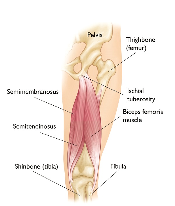
Posterior thigh muscles (hamstrings)
The hamstring muscles are a group of three long muscles which is located in the posterior compartment of the thigh, shaping up the surface anatomy of this region. The Hamstring group of muscles includes the biceps femoris, semitendinosus, and semimembranosus muscles.
The hamstrings muscles are closely related to each other as they share a common origin point, and they all attach to the proximal parts of the tibia and fibula. They are innervated by the tibial and common fibular (peroneal) divisions of the sciatic nerve (L4 to S3).
All the hamstring muscles cross the hip joint and knee joints and act upon them. The primary function of the hamstrings is to flex the knee joint and extend the hip joint, enabling some of the essential lower limb activities like walking, running, and climbing. The hamstrings also have an important stabilizing function; they are inactive when the body weight is equally distributed between both lower limbs in a standing position. However, when a person starts tilting forward, these muscles activate and counteract the tilting movement in order to stabilize the hip joint and prevent falling. Also, due to the location of their insertions, the hamstrings act together with the collateral ligaments to stabilize the knee joint.
Muscles on the back of the thigh name
The muscles of the posterior compartment of the thigh include the following muscles :
- Biceps femoris muscle: short head of biceps femoris muscle and a long head of biceps femoris muscle.
- Semitendinosus muscle
- Semimembranosus muscle
- The hamstring part of the adductor Magnus
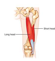
Biceps femoris muscle
The biceps femoris muscle is the most lateral muscle in the posterior compartment of the thigh and, as the term bicep means has two heads: the long head and the short head.
the long head of the biceps femoris and the short head of the biceps femoris muscle have different origins but join to form a palpable tendon on the lateral distal thigh inserting it onto the fibula head. The biceps femoris muscles’ long head protects the sciatic nerve as it passes from the gluteal region into the posterior compartment of the thigh.
Origin:
The long head of biceps femoris muscles originates from the inferomedial part of the ischial tuberosity
The Shorthead biceps femoris muscles originate from the lateral lip of the linea Aspera
Insertion:
Head of the fibula
Nerve supply:
The nerve supply of the long head of the biceps femoris is a tibial division of the sciatic nerve
The nerve supply of the Shorthead of the biceps femoris is a common fibular division of the sciatic nerve
Blood supply:
The inferior gluteal artery supplies the superior portion of the biceps femoris muscle
Perforating arteries of profunda femoris supply the majority of the biceps femoris muscle
Action:
Knee flexion
Hip extension
Lateral rotation at the hip and knee joint
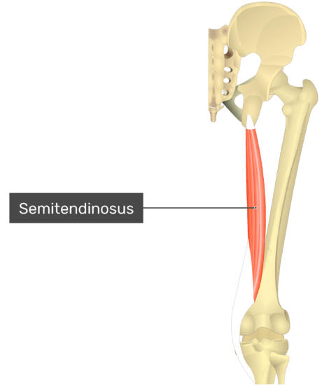
Semitendinosus muscle
The semitendinosus muscles are half tendinous. It lies medial to the biceps femoris and shares the same origin as its long head.
Semitendinosus lies superficial to semimembranosus muscles and forms its tendon approximately two-thirds of the way down the thigh. This tendon inserts to the medial surface of the tibia as part of the ‘pes anserinus’, which is also made up of the tendons of the sartorius and gracilis muscles.
Origin:
Semitendinosus muscles originate from the Inferomedial part of the ischial tuberosity
Insertion:
Semitendinosus muscles insert on the Medial surface of the tibia
Nerve supply:
Semitendinosus muscles nerve supply is a tibial division of the sciatic nerve
Blood supply:
The inferior gluteal artery supplies the superior portion of the Semitendinosus muscle
Perforating arteries of profunda femoris supply the majority of the Semitendinosus muscle
Action:
Knee flexion
Hip extension
Medial rotation of the hip joint
Medial rotation of the knee joint
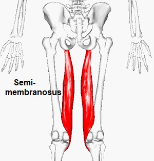
Semimembranosus muscle
The semimembranosus is named because of the flat membranous form of its proximal attachment. It also attaches to the ischial tuberosity but more superiorly compared to semitendinosus and the long head of the biceps femoris muscle.
Distally, the semimembranosus muscle tendon extends to contribute to the fascia and ligaments of the knee joint. It is the most medial muscle of the hamstrings and can be found underneath the semitendinosus muscles.
Origin:
The Semimembranosus muscle originates from the superolateral part of the ischial tuberosity
Insertion:
The Semimembranosus muscle inserted on the posterior surface of the medial tibial condyle
Nerve supply:
The Semimembranosus muscles nerve supply is the tibial division of the sciatic nerve
Blood supply:
The inferior gluteal artery supplies the superior portion of the Semimembranosus muscle
Perforating arteries of profunda femoris supply the majority of the Semimembranosus muscle
Action:
Knee flexion
Hip extension
Medially rotation of the hip joint
Medial rotation of the knee joint
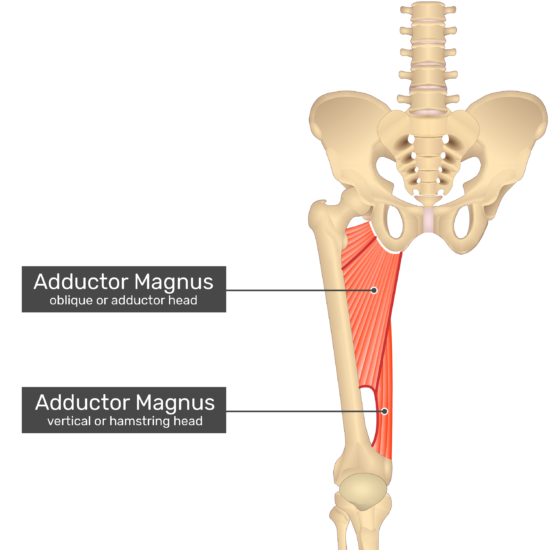
The hamstring part of the adductor Magnus
Origin:
The hamstring part of the adductor Magnus original from the lower lateral part of the ischial tuberosity, ramus of the ischium, and the lower part of the inferior ramus of the pubis
Insertion :
The hamstring part of the adductor Magnus inserted on the medial margin of the gluteal tuberosity, Linea Aspera, Medial supracondylar line, and Adductor tubercle
Nerve supply:
The nerve supply of the hamstring part of the adductor Magnus is the tibial part of the sciatic nerve
Blood supply:
The blood supply of the hamstring part of the adductor Magnus is the deep femoral artery
Action:
Extension of the hip joint
Flexion of the knee joint
Clinical Importance- Muscles of the back of the thigh:
Hamstring injuries
Injuries to the hamstrings muscles group are common for those competing in sports such as sprinting and football due to excessive stretching of the muscles. An injury is most likely to occur due to a sudden change in speed or direction. The level of injury can differ from a mild strain, where the muscles are excessively stretched, to a complete tear of the muscle itself.
In adults, the most common injured point is the muscle-tendon junction. Whereas, in adolescents, an avulsion fracture of the ischial tuberosity is more likely to occur. This is because the ischial apophysis (the site where the tendons attach) is the weakest part of the hamstrings muscles in this age group. In an avulsion fracture, the tendons of the muscle tear off and take a fragment of bone along with them.
MRI and ultrasound can be used to assess an injured hamstring, with MRI being able to provide more information about the level of the injury.
Use in surgery:
The distal semitendinosus tendon is one of the tendons that can be used in the surgical procedure of anterior cruciate ligament (ACL) reconstruction. In the anterior cruciate ligament (ACL) reconstruction procedure, a piece of it is used to replace the anterior cruciate ligament (ACL). The anterior cruciate ligament (ACL)is one of the four major ligaments in the knee, which also include the posterior cruciate ligament (PCL), medial collateral ligament (MCL), and lateral collateral ligament (LCL).