Obliquus capitis inferior muscle
Table of Contents
Obliquus capitis inferior muscle Anatomy
One of the four suboccipital muscles is the obliquus capitis inferior muscle. The largest muscle of the four is obliquus capitis inferior, which is grouped with the rectus capitis posterior major, rectus capitis posterior minor, and obliquus capitis superior muscles.
By supporting the atlantoaxial joint, obliquus capitis inferior helps to maintain posture and facilitate head and neck movements.
Origin and Insertion
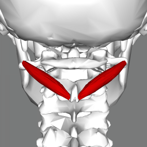
The only suboccipital group muscle that does not attach to the skull is the obliquus capitis inferior muscle. Rather, it spans the cervical vertebrae, first and second. Obliquus capitis inferior originates on the lateral side of the spinous process of the axis (second cervical vertebra), and it crosses superolaterally to attach to the posterior portion of the atlas’s transverse process (first cervical vertebra).
origin:
spinous process of the axis.
Insertion
lateral mass of atlas.
Nerve Supply
Suboccipital nerve.
The posterior ramus of spinal nerve C1, the suboccipital nerve, innervates obliquus capitis inferior, as it does all four muscles of the suboccipital group.
Blood Supply
The vertebral vein drains blood, whereas the vertebral artery supplies it. The occipital artery is a branch of the external carotid artery, and it descends deeply.
Actions
Rotation of head and neck.
Head extension at the atlantoaxial joint is produced by bilateral contraction of the obliquus capitis inferior. In contrast, a unilateral contraction causes the head to rotate to the ipsilateral side.
It should be noted that this muscle is a powerful rotator of the neck because of the oblique angle of its muscle fibers. The inferior obliquus capitis muscle is also crucial for maintaining good posture. It cooperates with the other suboccipital muscles to stabilize the atlantoaxial joint and, consequently, the head’s position during other bodily movements (e.g., getting up from a chair).
Relations
The posterior part of the neck contains the obliquus capitis inferior, which is situated deep to the semispinalis capitis, splenius capitis, and trapezius muscles. The rectus capitis posterior major and obliquus capitis superior muscles are part of the suboccipital triangle, which is an area of the neck.
This triangle’s inferolateral border is formed by the obliquus capitis inferior muscle.
FAQ
Head extension at the atlantoaxial joint is produced by bilateral contraction of the obliquus capitis inferior. In contrast, a unilateral contraction causes the head to rotate to the ipsilateral side. It should be noted that this muscle is a powerful rotator of the neck because of the oblique angle of its muscle fibers.
Of the two obliquus muscles, the larger one is the obliquus capitis inferior muscle. It starts on C2’s spinous process and travels laterally, then slightly superiorly, to attach itself to C1’s transverse process.
It is the sole capitis (Latin for “head”) muscle that is not attached to the skull, despite what its name might imply.
An uncommon disorder called cervical dystonia affects the obliquus capitis inferior muscle, which can lead to sudden, uncontrollable head jerks to the left or right, tilts from ear to shoulder, or oscillates back and forth. This excruciating condition may result in headaches, cramps in the neck, and straining.
Stretching between the first two cervical vertebrae, the obliquus capitis inferior (or musculus obliquus capitis inferior) is a tiny, slender muscle of the neck. It is situated in the neck’s posterior compartment. Therefore, one of the posterior neck muscles is the obliquus capitis inferior.
By twisting the client’s head and neck at the spinal joints to the left, the right-side obliquus capitis inferior (of the suboccipital group) is stretched. Remark: The atlanto-axial joint, which is located in the upper cervical spine, should be the primary target of the movement.

