Bed Sore
Table of Contents
Introduction:
Pressure ulcers, another name for bed sores, are wounds to the skin and underlying tissue brought on by applying pressure to the skin for an extended period of time. They frequently appear on bony regions such as the elbows, heels, hips, and tailbone, especially in people with restricted mobility. Prevention and management depend on proper care, frequent repositioning, and skin hygiene.
Bed sore:
Pressure ulcers are caused by forces that block the flow of blood to the skin:
- When too much pressure being exerted on the same area of the skin for too long, such as when someone lies in the same position all the time.
- When “shearing” forces cause the skin to bunch up in one area, such as when a person stays in a reclining position too long in either a bed or a chair.
Pressure ulcers can happen to:
- Newborns in incubators who are resting on lines or tubes.
- People with spinal cord injuries who have lost sensation and don’t feel uncomfortable sitting in the same position day after day—and therefore don’t realize that their skin is being affected.
- People who are bed bound and who are unable to change their position in bed.
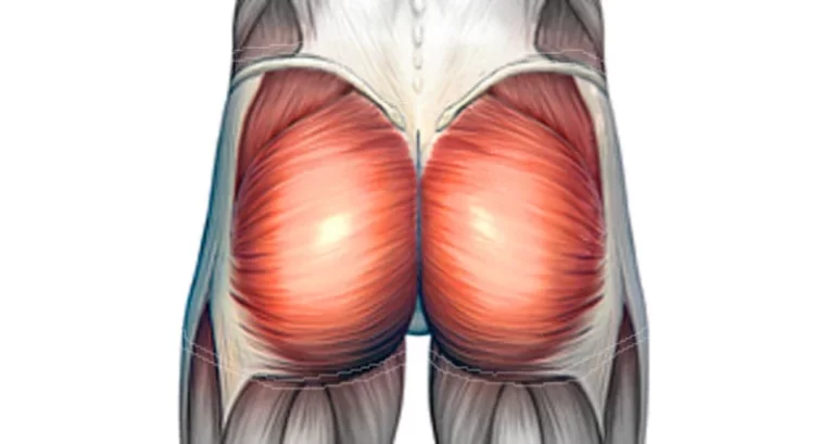
~Although pressure ulcers can develop anywhere on the body, they are more likely to occur on the buttocks of people who sit in a chair all day or in the heels, above the tailbone, and on the insides of the elbows of people who have to stay in bed all day.
~Pressure ulcers can result from friction injuries to the skin when a person is being pulled across a surface, such as being pulled across a sheet when the bed is being made or being pulled out of a wheelchair.
~Skin also can be injured by prolonged exposure to tape, urine, and feces, or it might be injured by tape removal. Although these injuries might look similar to a pressure ulcer, they often aren’t.
~However, this kind of wound is more likely to become a pressure ulcer if the skin is exposed to too much or too little moisture, scrubbing, or temperatures that are too cool or too warm.
The physical therapist will conduct a full evaluation, including staging the pressure ulcer based on a system developed by the National Pressure Ulcer Advisory Panel (NPUAP):
How is it diagnosed?
- Stage I ulcer – persistent reddening of the skin due to staying in the same position for too long. In darker skin, the skin might look purple. The skin might feel warmer to the touch than the surrounding skin.
- Stage II ulcer – shallow wound that goes only partially into the skin, usually caused by friction.
- Stage III ulcer – a deeper wound that goes through all of the layers of the skin, including fatty tissue under the skin.
- Stage IV ulcer – a wound that goes deeper than just the skin, down into the muscle, tendon, ligament, or bone; it might even expose nerves and blood vessels.
~These stages don’t necessarily tell how severe a pressure ulcer is, and ulcers don’t always progress from one stage to the next. What looks like a “simple” stage I ulcer might be a minor irritation of the skin—or might be hiding a large mass of injured tissue beneath.
~If there are blood-filled blisters, or purple or maroon areas of what appears to be uninjured skin, that could mean the tissue beneath the uninjured-looking skin is dead.
~On the other hand, there might be extensive death of tissue such as muscle, but the skin might not look injured at all. This is why a large ulcer might seem to appear “out of nowhere” within a matter of a few days.
~Muscle and other tissue near bone may be damaged by pressure before the skin breaks down, revealing a large wound extending to the bone. Detailed examination by a physical therapist or other health care provider is needed.
~Based on the examination, the physical therapist will decide whether any further testing or consultation with another health care provider is necessary. In some cases, surgery or the prescription of antibiotics by a physician may be necessary.
Physiotherapy management:-
~Wound care has been a part of physical therapist practice from its very beginnings. Based on the results of the physical therapist’s evaluation, including a review of the medical history and an examination of the wound, the therapist will select treatments, which may include caregiver training, strengthening exercises, wound care, improvements to the seat or bed, and coordination with other health care providers.
Train the Family and Caregivers in Repositioning:
~Because pressure ulcers are usually the result of prolonged contact of a body part with a bed or chair, repositioning or moving a person back and forth between a bed and a chair is needed to help pressure ulcers heal.
~Physical therapists are experts in positioning people on different surfaces, including beds, wheelchairs, toilets, bedside commodes, or other types of chairs and furniture as well as in moving a person from one surface to another (called “making transfers”).
~The physical therapist can train family members and caregivers to do positioning and transfers safely.
Improve Strength:-
~In many cases, pressure ulcers are the result of people being too weak to change their own position.
~The physical therapist will develop an exercise program so that the individual with pressure ulcers can help perform repositioning and transfers, in turn helping to prevent injury to caregivers.
~In some cases, a person with a pressure ulcer may regain the ability to do repositioning without any help.
Care for Wounds:-
~The physical therapist is trained in wound care and knows how to remove “nonviable,” dead tissue from a wound (“debridement”). The therapist may use sharp instruments such as scissors, scalpels, or curettes.
~Extensive debridement that requires cutting of surrounding healthy tissue or that requires general anesthesia would be performed by a surgeon.
~Pressure ulcers require appropriate dressings to aid healing. Dressings include a suitable material to cover wounds and to fill deeper wounds. The physical therapist determines the type of dressing to use and how frequently it must be changed.
~In most cases, caregivers can be trained by the physical therapist to clean wounds and to change dressings. As a pressure ulcer heals, the ulcer likely will change, and so will the treatment.
~The physical therapist will evaluate the wound with each visit and determine the type of dressing to use and how often it should be changed.
~The physical therapist will discuss what to expect in terms of normal wound healing, signs to look for that might indicate a problem between visits, and when to contact a physician or an emergency department.
Improve Sitting and Lying Surfaces:-
~The surface on which a person lies or sits may contribute to pressure ulcers. The physical therapist will examine the types of surfaces being used and, if necessary, will make recommendations for a different surface.
~Recommended support surfaces may include specialized beds, mattresses, or cushions made of materials that decrease the amount of pressure on vulnerable parts of the body.
Communicate With Other Health Care Providers:-
~A number of health care providers may be involved in the care of a person with a pressure ulcer. A physical therapist will communicate and coordinate care with physicians, nurses, dietitians, and others to ensure as much healing as possible and to prevent new ulcers.
*Ultrasound therapy:-
~Us can increase tissue temperature and it includes acceleration of metabolic rate, reduction or control of pain and muscle spasm, increase circulation and increase soft tissue extensibility.
~it heats smaller and deeper areas than most superficial area.US heats tissue with high US absorption coefficient -with high collagen content like tendon ligament joint capsule but not for fat with water content.
~US is not ideal for muscle heating because of low absorption but is very effective in healing scar in muscle areas because of increased collagen content.
~application of ultrasound stimulates cell activity and accelerates the inflammatory process.
~the skin repair and wound contraction will be accelerated.
~US stimulates collagen secretion and has effect on elastin properties which strengthen scar tissue.
~procedure is done by covering the wound with a hydrogel and delivering US by a hand held applicator.
~another option is to apply US transmission gel over the peri-wound area and treat from this region instead of the wound bed.
~the parameters that have been found to be effective for healing wound is 20% duty cycle,0.8-1.0 W/cm2 intensity,3Mhz frequency, for 5-10 minutes.
~treatment duration depends on the area of the wound.

Electrical stimulation:-
~electrical stimulation is effective in facilitating healing in both acute and chronic wounds.
~it is used to eliminate bacterial load,promote granulation,reduce inflammation,edema,reduce wound-related pain.
~electric stimulation has a galvanotoxic effect on the cell needed for healing.
~by using high volt pulsed current (HVPC)directly in wound can create these changes -attraction of neutrophils,macrophages, and epidermal cells which facilitate debridement and reepithelialization.
*Methods of application:-
~direct method of application:-it includes an ES unit treatment and non treatment electrodes and a saline soacked gauze or hydrogel dressing over wound bed to enhance electrical conductivity.
~indirect method of application:-here electrodes are placed around the periwound skin using gel.
Radiant gel:-
~infrared radiation increases local wound and skin temperature facilitating metabolic rate and improving circulation to the wound site.
~this technique is effective in treating chronic wounds even in the presence of vascular compromise.
~normothermia can be accomplished by warm up wound thrapy system which includes,delivering moist heat through a non contact dressing.
~using a warming card which is placed in sleeve on top of the sterile wound cover giving warmth up to 38 degree c.

*Negative pressure wound therapy(NPWT):-
~NPWT is a wound healing technique used to facilitate wound closure in acute surgical and challenging slow healing wounds.
~VAC or vaccum assisted closure is the device used to provide negative pressure treatment.
~an open cell foam dressing is placed in the wound and a suction tube is connected from the foam to the portable pump,an air tight seal is created over the foam and suction tube with a film.
~a controlled amount of negative pressure (subatmospheic)is applied through the foam to the wound bed.
~for the first few days 48 hrs pressure applied continuously via a portable pump, after the withdrawal of a significant amount of wound fluids it is done intermittently .
~the foam is changed every 12 hrs.(infected wounds).


Short wave Diathermy:-
~PSWD and CSWD have been used to treat chronic open wounds.
~it provides radio waves to produce thermal and non thermal effect by facilitating one pahse of healing to next.
~PSWD heats superficial tissues and CSWD heats deep muscle and joint tissue.
~it increases fibroblast proliferation,collagen formation and tissue perfusion.
~treatment is delivered usually without touching the skin,but with newer units pad can be placed over the wound dressing, compression garments etc.
Ultraviloet radiation:-
~it is a form of energy between x-ray and visible light
~it is divided into wavelength and bands.
~three bands useful for human skin are UVA,UVB AND UVC.
~it has bactericidal effects and it increases blood flow, enhances granulation tissue formation, stimulation of vitamin D.
~procedue is done on a clean wound with dressing removed using UVB or UVC lamp.
~treatment distance dosage frequency will vary on the status of the wound.
*Hyperbaric oxygen therapy:-
~HBO delivers 100% o2 to an individual who rests inside a sealed chamber at a pressure greater than atmosphere(full body chamber).
~it increses amount of o2 available for cell metabolism increase o2 in hypoxic tissue.
~topical hyperbaric o2 therapy THBO is used now a days instead of full body chamber,localized limb chambers are used, so THBO delivered o2 directly to the surfcae of the wound trough a portable unit.
~it is also used in combination therapy along with stimulation or with cold laser.


Compression therapy:-
~the concept of compression therapy is based on a simple and efficient mechanical principle consisting of applying an elastic garment around an area of the body to control edema.
~edema not only inhibits wound healing by affecting perfusion of the tissue but also inactivates the ability of the skin to manage bacteria.
~it should apply as soon as signs of swelling appear when leg wounds are present.
Elevation:-
~it is not a compression technique but used to reduce some types of swelling (mild acute swelling)and is a precursor to compression.
~proper positioning and active ROM exercise should teach the patient to incorporate with other means of swelling controlling techniques like compression etc.
Four-layer bandage system:-
~four layer bandaging is a high compression bandaging system (sub bandage pressure 35-40 mm hg at the ankle)that incorporates elastic layers to achieve a sustained level of compression over time.
~since the development of the four-layer system over 15 years ago.
~the four-layer bandage system is primarily used in the treatment of venous ulceration and achieves healing in patients with both deep, superficial, and combined venous incompetence.
~four layer bandaging can also be used to prevent recurrence in patients who are unable to wear elastic stockings.
~the short stretch, an elastic effect noted in four-layer bandaging has made this a useful treatment.
Indications:-
*primary uses:-
~treatment of venous ulceration
~prevention of ulcer recurrence if hosiery is not tolerated.
~symptomatic relief of superficial thrombophlebitis.
*Other use:-
~traumatic wounds with local edema, for example, pretibial lacerations.
~venous/lymphatic disorders.
~ulceration of mixed aetiology with an oedematous component.
*Contraindications:-
~patients with heart failure should not receive high compression therapy.
~in this instance high compression will redistribute blood towards the center of the body, thereby increasing the pre-load of the heart and possibly causing further overload and death.
~patients with severe obliterative arteriosclerosis should not receive compression therapy.
*Application:-
~Layer-1:orthopedic wool:-orthopedic wool provides a layer of padding that protects areas at risk of high pressure.
~layer-2:crepe bandage:-this is the least effective layer as it simply adds extra absorbency and smooths down the orthopedic layer prior to the application of the two outer compression bandages.
~layer-3:elastic extensible bandage:-it is a highly extensible bandage that provides a sub bandage pressure of approximately 17 mm hg when applied at 50% overlap using a figure of eight technique.
~layer-4:-elastic cohesive bandage:-a frequent misconception is that the outer cohesive layer within the four layer system is there simply to maintain bandage position,in fact thhis layer provides the higher lavel of compression (sub bandage pressure approximately 23 mmHG).

*Long and short stretch bandages:-
~this both bandages are used to control edema nd provide compression to support the lymphatic system.
~long stretch bandages provide a high resting pressure means they constrict when the wearer is resting.
~they do not provide significant working pressure.they are readily available and easy to wear.
~short stretch bandages provide low resting pressure but provide high working pressure.
~they are less stretchy, provide a rigid appearance after application and this makes them more appropriate for edema treatment.
~working pressure increases the work of muscle like pumping activity and lower resting pressure makes bandage more tolerable.
~it needs special training to apply like no: layers, age condition and tension of the bandage, etc.
*Lymphedema bandage:-
~this is a highly specialized bandage with multiple layers of padding materials and a short stretch bandage that provides support to the lymph edematous body part.
~it provides support to the tissues with elasticity loss and facilitates a mild tissue pressure to empty the lymph vessels.
~it is applied to the head and neck,chest,abdomen,genital area and back.


*Compression garments:-
~it is widely used by clients all over the world ,it is designed to venous blood flow is Les.
~now it is designed to manage burns surgical scars to provide support to venous circulation ant to prevent reaccumulations of fluids it is not used as a treatment to remove excess fluids.
~another one is quilted garment which provide compression which is used by person who can not apply support garment and whose skin is fragile.
~venous return and lymphatic drainage is attained by altering the stitching channels.


*Guidlines for compression bandaging:-
~arterial wound:-no compression or very light long stretch bandage in 12-25 mmHG is used.
~venous wounds:-compression is essential,short stretch bandage with high working preassure 40 mmHG.
~neuropathic wounds:-if no arterial involvement compression with short stretch wrap.
~lymphedema:-short stretch compression wrap untle limb reduction then modarate to high compression 20-30 mm HG 30-40 MMHG.
~edema same as lymphedema short stretch compession 23 hours/day.
*wound dressing:-
~a dressing is an adjunct used by a person for application to a wound to promote healing or to prevent further harm. a dressing is designed to be in direct contact with wound,which makes it different from a bandage.
~choosing appropriate dressing should be on the basis of wound and periwound tissue.a product that preserves wound hydration limit fluid loss is ideal.
~in moist wound dressing the following wound characteristics must be considered.
*infection-present/absent
*necrosis-remove/not
*drainage-dry,adequate or excessive
*granulation-present/not
*epithelielization-present/not
*periwound area-intact/at risk
*odor-minimal /need reduction
*primary dressing-that applied directly to the wound
*secondary dressing-that applied over primary one