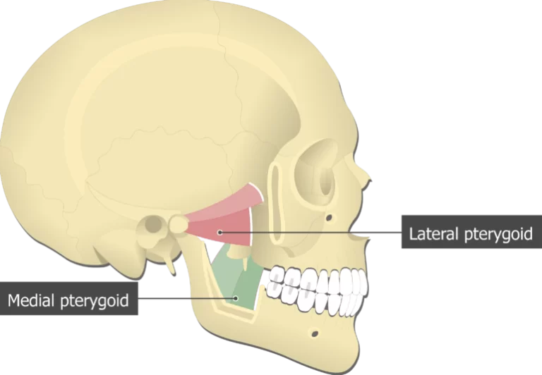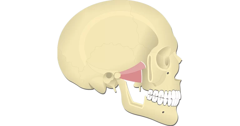
Lateral pterygoid Muscle
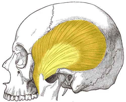
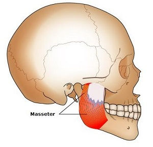
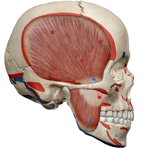
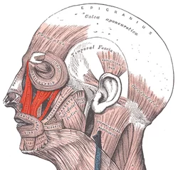
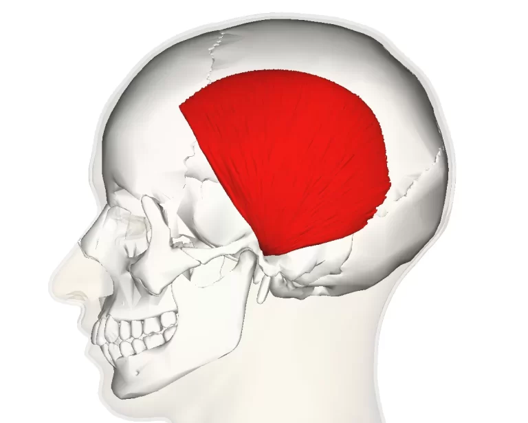
Introduction The lateral pterygoid muscle has two heads the upper head and the lower head.The lateral pterygoid is the fan-shaped muscle located laterally in the infratemporal fossa of the skull. It is one of the four mastication muscles, along with the medial pterygoid muscle, temporalis, and masseter muscles. All these mastication muscles act upon the…