
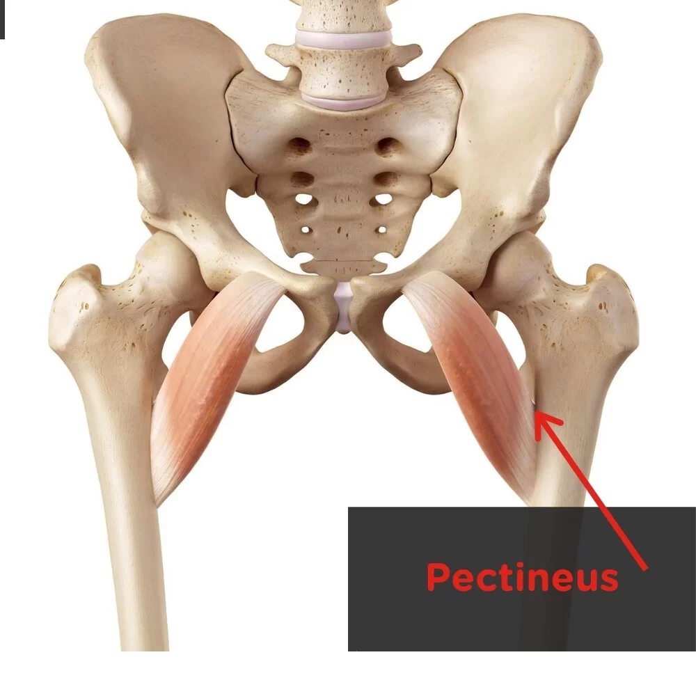
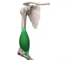
Pectineus Muscle
Pectineus Muscle is a flat, quadrangular muscle, situated at the anterior (front) part of the upper and medial (inner) aspect of the thigh.
Together with the other adductor muscles, the pectineus muscle adducts the thigh or brings the leg closer to the midline of the body. The pectineus muscle begins from the pubic bone’s pectineal line and ends at the femur, or thigh bone, pectineal line.
It is the hip’s most anterior adductor, innervated by the obturator nerve on the muscle’s posterior surface and the femoral nerve on its anterior surface. Together with the femoral and obturator arteries, the medial circumflex femoral artery provides the majority of the blood flow to the pectineus muscle.
The pectineus muscle is situated laterally to the medial circumflex femoral vein and artery, as well as the psoas major muscle. A thick layer of connective tissue called the fascia lata covers the anterior surface of the pectineus muscle, separating it from the femoral artery, femoral vein, and great saphenous vein that all pass through the femoral triangle.
Origin:
Pectineal line of the pubis and a narrow area of the superior pubic ramus below it.
Insertion:
A vertical line from the lesser trochanter to the linea Aspera.
Nerve supply:
Anterior division of the femoral nerve (L2, L3).
Action:
Assists with flexion and adduction of the thigh.
Flexion at the hip joint results from muscular contraction when the lower limb is in its anatomical position. This flexion can extend to the point where the thigh and hip joint are at a 45-degree angle.
Thigh adduction results from the tightened muscle fibres pulling the thigh towards the midline at that location due to the fibres’ angulation. Leg crossing at the knee or ankle is an illustration of a sequential movement using both pectineus muscle movements. Only hip flexion facilitates the carry-through phase of the gait cycle when combined with the psoas major, iliacus, rectus femoris, and sartorius.
Table of Contents
Relations
The muscle is situated medially to the adductor longus and in the same plane. Laterally, it is associated with the medial circumflex femoral vein and artery as well as the psoas major muscle.
Together with the adductor longus, the anterior aspect of the pectineus forms the medial portion of the femoral triangle floor, while the iliacus and psoas major complete the lateral side. The dense layer of fascia lata that covers the pectineus surface divides it from the femoral artery, femoral vein, and great saphenous vein that passes through the femoral triangle.
The adductor externus, adductor brevis, and adductor magnus muscles, as well as the anterior branch of the obturator nerve, are located posterior to the pectineus. It should be noted that the anterior and posterior thigh compartments are peripherally linked by the fascia lata. The lateral and medial intermuscular septa, which connect to the femur, divide these two internally. Due to its shared purpose and the fact that all of its component muscles are innervated, the adductor (medial) compartment, to which the pectineus belongs, is still classified separately from the anterior and posterior compartments despite lacking a distinct fascial boundary.
Strengthening exercise:
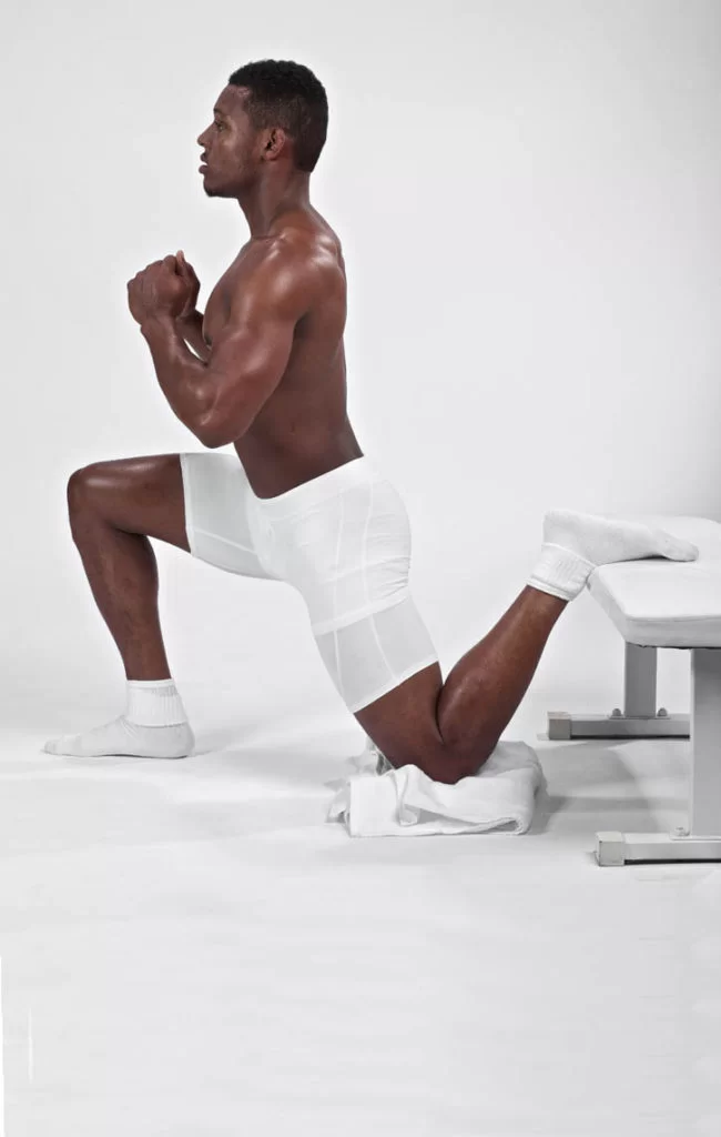
1. Standing hip flexion exercise:
Stand facing a bench or other elevated surface, such as a secured and stable chair. Distance yourself from it approximately the length of your leg. With your hands on your hips, place one foot on the bench or elevated surface. Bend your raised leg slowly, causing you to lunge forward, while keeping your torso straight. Push your hips slightly forward and hold the stretch for at least two seconds, but you can hold for longer as your strength increases. Perform ten repetitions on one side, then repeat with the other leg.
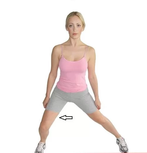
2. Lateral Lunges:
Begin by standing with your feet a little less than shoulder-width apart. Place your hands on your hips, or out to the side to help with balance. Step your right foot out to your right side, and into a squat position to the one side. Go as far down as you are comfortable doing, but not past the point of where the thigh is parallel to the ground. Gently push with your right foot to stand back up again and return the right foot to the starting position. Keep your core stable by contracting your abdominal muscles while performing this exercise. Perform this exercise four times on each side, increasing the number of repetitions and sets as your strength increases.
Stretching exercise:
Kneeling Lunge and Overhead Stretch
Place your right hand on your right knee, extend your left arm overhead, and bring the arm slightly across your body to the right. Feel the stretch in your pectineus and upper body (shoulder and lat muscles) as you lunge slightly forward with your right leg.
Related pathology:
The pectineus muscle can become injured by overstretching; specifically, by stretching a leg or legs too far out to the side or front of the body. Pectineus injuries can also be caused by rapid movements like kicking or sprinting, changing directions too quickly while running, or even sitting with a leg crossed for too long.
What causes pectineus muscle pain?
A damaged pectineus muscle is the most frequent cause of pectineus pain. Stretching the legs too far out to the side or front of the torso may result in injury to the pectineus muscle.
Overstretching of the muscle can also occur from fast motions like kicking, sprinting, abrupt direction changes when jogging, or prolonged sitting with the legs crossed. Bruising, swelling, soreness, and stiffness in the inner thigh are further signs of a pectineus injury.
Treatment
When treating a pectinus muscle injury, the patient should protect the injured area from movements and objects that could aggravate the injury further; limit activities that use the pectinus muscle, such as walking and running, to give the muscle time to heal; and apply ice to the injury to reduce swelling and pain. Every few hours, ice should be done for 15 to 20 minutes.
FAQ
A pulled or strained muscle is the most frequent cause of pectineus pain. The pectineus can be injured by jogging, skating, working out weary muscles, or even just sitting with your legs crossed for an extended period of time. Pectineus injuries can be treated with time, ice, physical therapy, and rest.
Axial pictures make it simple to identify the pectineus muscle because it forms the floor of the femoral triangle across which the femoral neurovascular bundle flows anteriorly. The muscles known as the adductor longus and adductor brevis have a broad insertion onto the femur.
The Gluteus Maximus is the antagonist of the Pectineus. A muscle that contracts when the other relaxes is known as an antagonist muscle. Thus, the Gluteus Maximus relaxes as the Pectineus contracts.
Treatment for pectineus pain typically involves rest, ice, and anti-inflammatory medications to reduce inflammation and alleviate discomfort. Physical therapy exercises may be recommended to strengthen and stretch the muscles. In severe cases, a healthcare professional might suggest corticosteroid injections or other interventions.
Pain is the most typical sign of a pectineus muscle injury. Additional symptoms might be stiffness, discomfort, edoema, and bruises.
5 Comments