The Cerebrum
Table of Contents
Introduction:
~the cerebrum is made of two cerebral hemispheres which are incompletely separated from each other by the median longitudinal fissure.
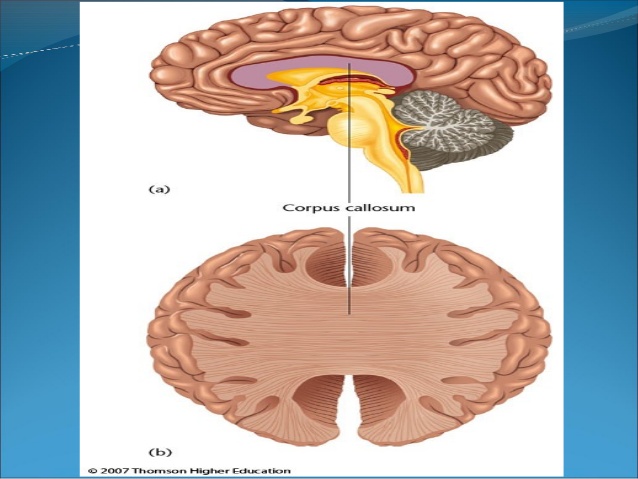
~the two hemispheres are connected to each other across the median plane by the corpus callosum. each hemisphere contains a cavity, called the lateral ventricle.
External feautures:
three surfaces:
1. the superolateral surface is convex and is related to the cranial vault.
2. the medial surface is flat and vertical.it is separated from the corresponding surface of the opposite hemisphere by the falx cerebri and the longitudinal fissure.
3. the inferior surface is irregular.it is divided into an anterior part, the orbital surface, and a posterior part,the tntorial surface .the two parts are separated by a deep cleft called the stem of the lateral sulcus.
four borders:
1.the superomedial border separates the superolateral surface from the medial surface.
2. the inferolateral border separates the superolateral surface from the inferior surface. the anterior part of this border is called the superciliary border. there is a depression on the inferolateral border situated about 5 cm in front of the occipital pole, it is called the preoccipital notch.
3.the medial orbital border separates the medial surface from the orbital surface.
4.the medial occipital border separates the medial surface from the tentorial surface.
three poles:
1.frontal pole:-at the anterior end
2.occipital pole:-at the posterior end
3.temporal pole:-at the anterior end of the temporal lobe.
Sulcai and gyri:
~the surface of the each hemisphere is covered by a thin layer of grey matter called the cerebral cortex.the cortex is folded in a complex manner so that there are a number of raised areas (gyri),that are separated by depressions(sulcai)the pattern of sulcai and gyri will be studied in subsequent sections.
Cerebral hemisphere:
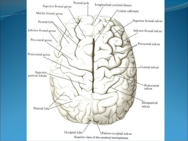
~general appearance:
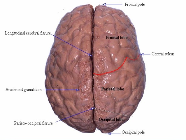
~separated by a deep midline saggital fissure:-longitudinal cerebral fissure.
~the fissure contains falx cerebri and the anterior cerebral arteries.
~in the depth of the fissure ,the corpus callosum connects the hemisphere across the midline.
~gyri:-the folds of the surface of hemisphere.
~sulcai:-the fissures separates the gyri.
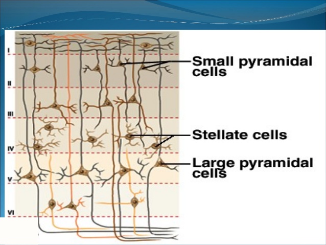
Cerebral cortex:
~surface layer of grey matter-3 mm thick.
~neocortex(six layered tissue)
~newest part of the cortex (paleocortex & archiocortex)
~layers vary in thickness in different regions of brain.
~2 types of cells:-
~stellate cells :-have dendrites projecting in all directions.
~pyramidal cells:-have an axon that passes out of the area.
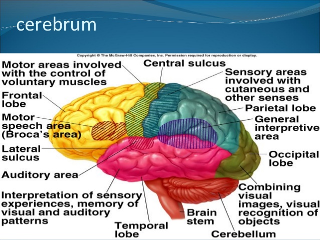
cerebral cortex-functions:
~the is particularly well developed in humans.
~in responsible for many higher brain functions.
~including manual dexterity (eg.to move the fingers individually so as to play piano).
~conscious,discriminative aspects of sensation.
~congnitive activity,inculding language,reasoning and
~many other aspects of learning and memory.
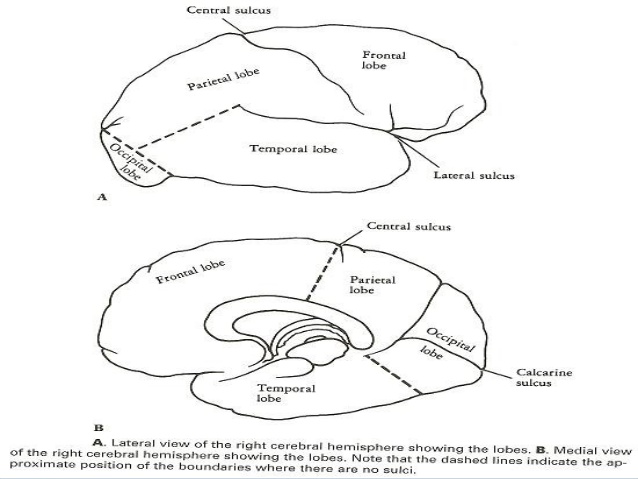
Lobes of cerebral hemisphere:
~each cerebral hemisphere is divided into four lobes-central,parieto-occipital,lateral and calcarine sulcai.
~lobes are named according to cranial bones under which they lie:-
~lobes are:-
~frontal
~parietal
~temporal
~parietal
~temporal
~occipital
The cerebral cortex (consists of sixes lobes on each side)
~frontal
~parietal
~temporal
~occipital
~insular
~limbic
~the underlying white matter
~the basal ganglia:-a complex of deep gray matter masses.
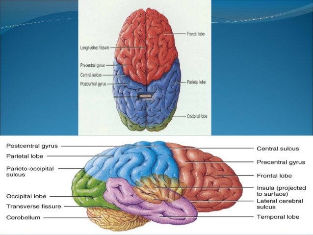
*The main sulci include:
1.central sulcus:
2.Lateral sulcus:
~indents the superior medial border of the hemisphere,1-cm behind the mid point.
~it runs downward,forward and toward the lateral sulcus across the lateral aspect of the hemisphere.
~the central sulcus is the only sulcus that indents the superior medial border.
~deep cleft on the inferior and lateral surfaces of the cerebral hemisphere.
~it consists of short stem and three rami.
3.Parieto occipital sulcus:
~begins on the superior medial border of the hemisphere,about 5 cmanterior to the occipital pole.
~it passes downward and anteriorly on the medial surface to meet the calcarine sulcus.
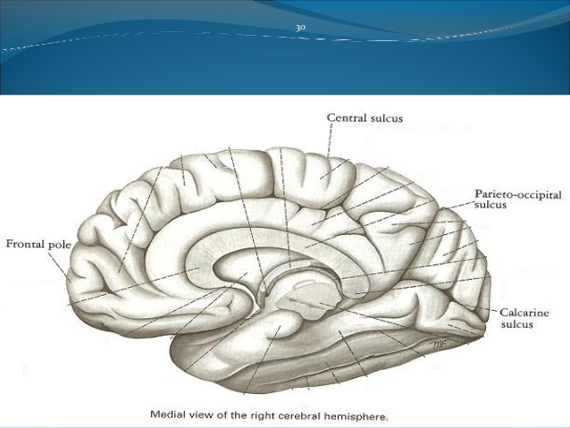
4.Calcarine sulcus:
~found on the medial surface of the hemisphere.
~it begins under the posterior end of the corpus callosum.
~it ascends upward and backward to reach the occipital pole.
Surfaces of cerebral hemisphere:
~superolateral surface
~inferior surface
~medial surface
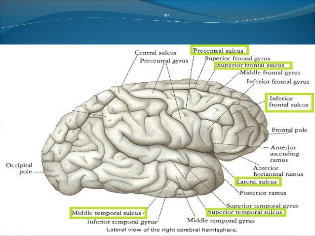
Superolateral surface:
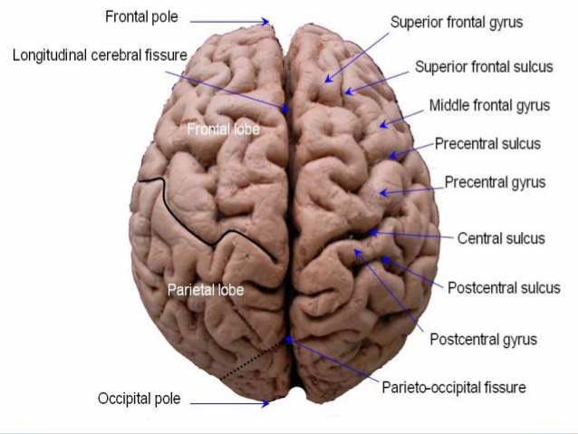
~Frontal lobe:-anterior to central sulcus and superior to lateral sulcus.
~superlateral surface of frontal lobe is devided by three sulci into four gyri.
~precentral sulcus and gyrus.
~superior and inferior frontal sulci.
~superior ,meddle and inferior frontal gyri.
Cerebral cortex:
~frontal lobe:-anterior portion of each cerebral hemisphere.
~precentral gyri:-contains upper motor neurons.
~involved in upper motor control.
~body regions with the greatest number of motor innervation are represented by the largest areas of motor cortex.
Superolateral surface:
~temporal lobe:-inferior to lateral sulcus.
~two sulci and three gyri.
~occipital lobe:-small area behind the parieto-occipital sulcus.
~gray matter:-cerebral cortex
~Lateral ventricles.
~basal nuclei-masses of gray matter.
~white matter:-nerve fibers
Cerebral cortex:
Temporal:
~contains auditory centres that receive sensory fibers from cochlea.
~interpretation and association of auditory and visual information.
Occipital:
~primary area responsible for vision and coordination of eye movements.
Insula:
~implicated in memory encoding.
~integration of sensory information with visceral responses.
~coordinate cardiovascular response to stress.
Superolateral surface:
~Parietal lobe:-posterior to cental sulcus and superior to lateral sulcus ,extends upto parieto-occipital sulcus.
~two sulci and three gyri.
Parietal lobe:
~primary area responsible for preception of somatesthetic sensation.
~body regions with the highest densities of receptors are represented by the largest area of the sensory cortex.
Medial and inferior surface:
~important areas are:
~corpus callosum
~cingulate sulcus and gyrus
~paracental lobule
~precuneus and cuneus
~occipito-temporal,collateral and calcarine sulcus.
~parahippocampal,medial and lateral occipitotemporal gyrus and uncus.
~olfactory sulcus,gyrus rectus and orbital gyri.
Internal structure of cerebral hemisphere:
~gray matter:-cerebral cortex
~Lateral ventricles.
~basal nuclei-masses of gray matter.
~white matter:-nerve fibers
Lateral ventricle:
~two lateral ventricles:-one is in each cerebral hemisphere.
~it communicates with the third ventricle through interventricular foramen.
~C-shaped
~body-lies in the parietal lobe
~anterior horn-frontal lobe
~posterior horn:-occipital lobe
~inferior horn:-temporal lobe
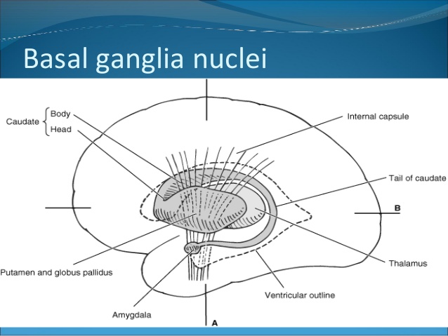
Basal ganglia:
~anatomically,the basal ganglia include
~the caudate nucleus
~the putamen,and the globus pallidus(together they are called the corpus straitum)
~functionally,the basal ganglia and their interconnections and neurotransmitters from the extrapyramidal system.
Basal nuclei:
~corpus straitum
~amygdaloid nucleus
~claustrum
White matter:
~composed of mylinated nerve fibers
~classified into three groups:-
~commissural fibers
~association fibers
~projection fibers
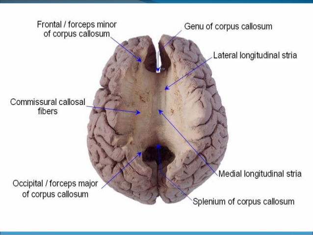
Corpus callosum:
~largest commissure
~parts:-rostrum
body
genu
splenium
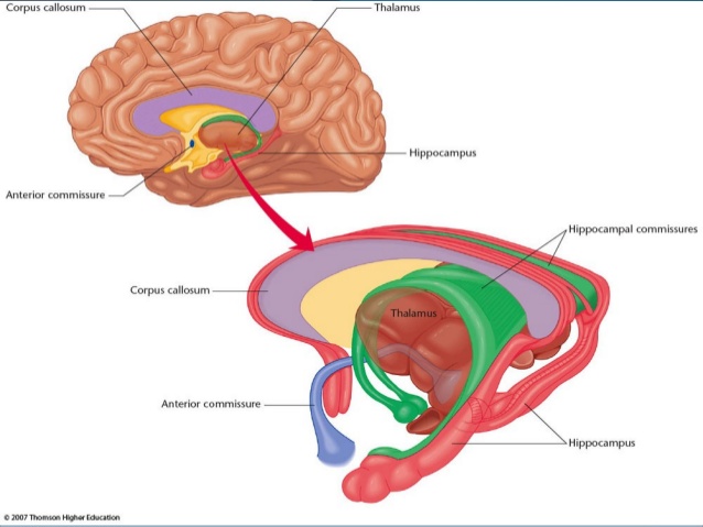
Commissural fibers:
~connect corresponding regions of two hemispheres.
~they are:-corpus callosum
anterior commissure
posterior commissure
fornix
habenular commissure
hippocampal commissure
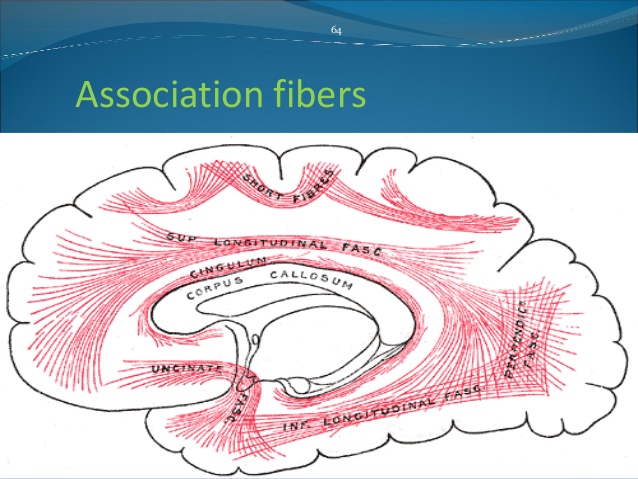
Association fibers:
~connects various cortical regions within the same hemisphere.
~they are:-
~short association fibers-connect adjacent gyri
~long association fibers-collected into named bundles.they are:-
-uncinate fascicullus
-cingulum
-superior longitudinal fascicullus
-inferior longitudinal fascicullus
~fronto-occipital fascicullus
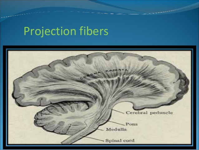
Projection fibers:
~afferent and efferent nerve fibers passing to and from the brainstem to the entire cerebral cortex.
~they are:-
-internal capsule
-corona radiata
-optic radiation
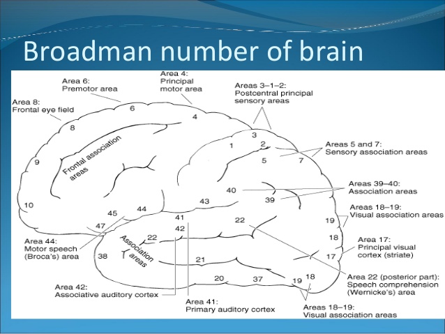
Areas of cerebrum:
~broadmann numbers to identify functions down to individual sulci.
~question localisation now that we know more about connectionism and we have a more dynamic view of how the brain works.
Cerebrovascular dieases:
Stroke:
~a cerebrovascular accident defined as an abrupt onset of a neurological deficit that is attributable to a focal vascular dieases.
two types:
1.Ischemic stroke:-
1.thrombotic stroke
2.embolic stroke
2.hemorrhagic stroke:-
1.intracerebral hemorrhage
2.subarachnoid hemorrhage
Ischemic stroke:-
~lacunar 20%
~perforating arteries obstruction,microatheromas,microembolis
~basal ganglia
~very isolated lesion:-pure motor or pure sensory stroke
~intracranial atherosclerosis 50%
~hemodynamic:-more common in the internal carotid artery
Hemorrhagic stroke:
~intracerebral haemorrhage:-uncontrolled hypertension with greater bleeding in the substances of brain.
~15% of stroke
~classic symptoms:-sudden onset of headache,vomiting and elevated BP.
~focal neurological deficit progress over minutes.
~may present with agitation and lethergy,then progress to stupor or coma.
Subarachnoid hemorrhage:
~aneurysm(>40 yrs)
~av malformation(<40 yrs)
~greater in between the pia and arachnoid matter.
~bleeding into the space between the inner and the middle layer of the tissue covering the brain.
~cause sudden ,severe headache,often followed by a brief episode of unconsciousness.
Symptoms of stroke:
~loss of sensory and motor function on one side of the body.
~change in vision,gait or ability to speak or understand
~sudden severe headache
~dizziness
~confusion
~loss of balance or coordination
~nausea/vomiting
~seizure
~painful stiff neck
Management:-
1.Ischemic stroke:
~BP management
~recognize sings of inceresed ICP
~non cardio embolic:-antiplatelet or combination,neurospecialist for posterior circulation stroke,if cerebral infarct refer to neurosurgery.
Hemorrhagic stroke:
~medicle compression:-mennitol,furosemide,hypertonic saline
~long term strict BP control
~neurosurgical control
~ICP monitoring
~goal:-reduce mortality
Prevention:
~diet(low fat,high fiber)
~quit smoking and alcohol intake
~control diabetes
~maintain healthy weight
~exercise
Crebrovascular terms:
~Hemiplegia
~Hemiparesis
~dysarthria
~aphasia:-expressive aphasia,receptive aphasia
~hemianopsia