
The spinal cord
Table of Contents
What is the spinal cord?
~the spinal cord is the lower elongated, cylindrical part of the CNS. It occupies the upper two-thirds of the vertebral canal.it extends from the level of the upper border of the atlas to the lower border of vertebra L1 or the upper border of vertebra L2.
~it is about 45 cm long. the lower end is conical and is called the Conu medullaris.
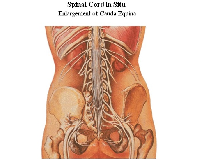
~the apex of the conus is continued down as the filum terminale.
~along its length,the cord presents two thickenings,the cervical and lumbar enlargements,which give rise to large nerves for the limbs.
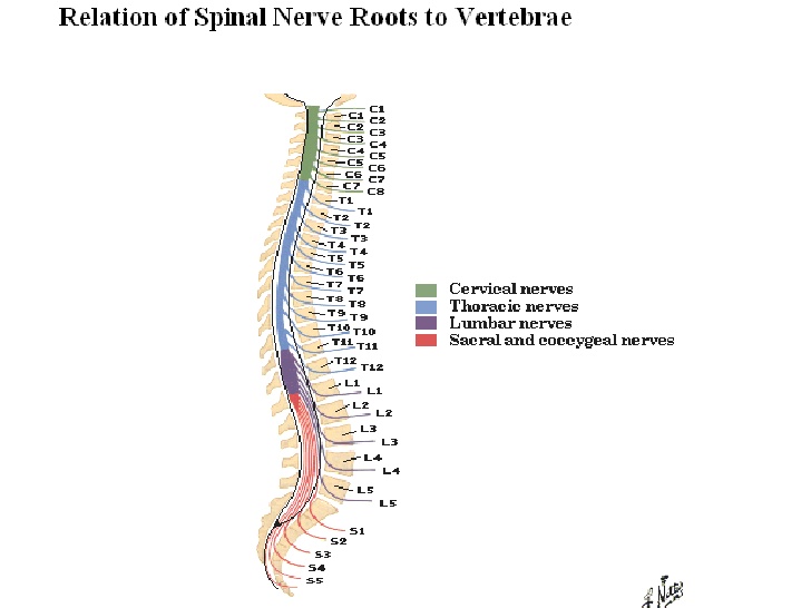
~the spinal cord gives off 31 pairs of spinal nerves.
Meningeal layers around the spinal cord:-
~duramater S2
~arachnoidmater S2
~piamater L1
~filum terminale
~linea splendens
~ligamenta denticulata
spinal nerves & spinal segments:-
~gives off 31 pairs of nerves
~C8 T12 L5 S5 CO1
~Cervical & lumbar enlargements
~cauda equina
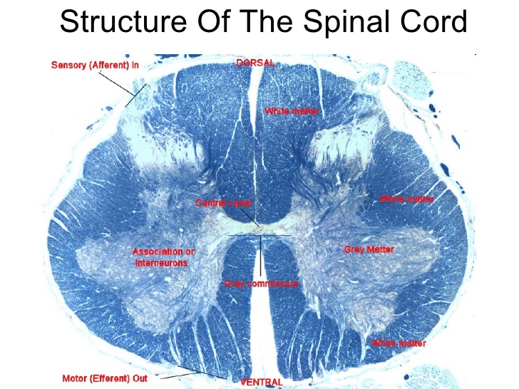
Gray mater:-
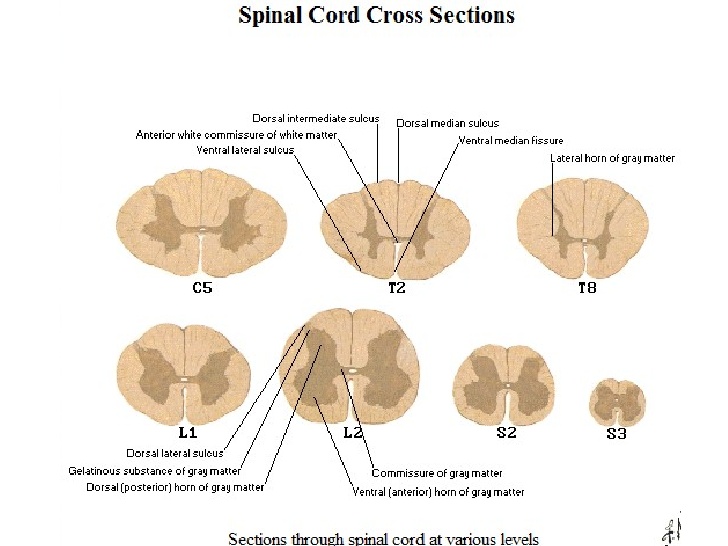
~H-shaped pillar with anterior & posterior gray mater horns.
~united by gray commisure containing the central canal.
~lateral gray column (horn)present in thoracic & upper lumbar segments.
~amount of gray mater related to the amount of muscle innervated.
~consists of nerve cells ,neuroglia,blood vessels.
nerve cells in the anterior gray columns:-
~large & multipolar
~axons pass out in the anterior nerve roots as efferents.
~smaller nerve cells are multipolar.
~axons pass out in anterior roots as effrents.
nerve cells in the posterior gray columns.
~4 nerve cell groups:-
~substania gelatinosa:-
~situated at the apex.
~throughout the length of spinal cord.
~composed mainly of golgi type || neurons.
~recevies afferent fibers concerning with pain,tempreture & touch from posterior root.
Nucleus proprius:-
~anterior to substania gelatinosa.
~present throughout the length of spinal cord.
~main bulk of cells in posterior gray column.
~recevies fibers from posterior white column that are assoc with proprioception ,2-point discrimination,vibration.
Nucleus dorasalis:-
~base of posterior column
~C8-L3/L4
~associated with proprioceptive endings.(neuromuscular spindles & tendon spindles)
Visceral afferent nucleus:-
~lateral to nucleus dorsalis.
~T1-L3
~receives visceral afferent info
Nerve cells in the lateral gray columns.
~formed by the intermediolateral group of cells.
~T1-L2/L3
~cells give rise to preganglionic sympathetic fibers.
~in S2,S3,S4 ,they give rise to preganglionic parasympathtic fibers.
The gray commissure & central canal:-
~connects the gray on each side.
~central canal in the centre
~posterior gray commissure
~anterior gray commissure
~cental canal present throughout
~superiorly continous with the central canal of medulla oblongata
~inferiorly,expands as terminal ventricle
~terminates within the root of filum terminale.
White matter:-
~devided into:-
~anterior white column
~posterior white coumn
~lateral white column
~consists of nerve fibers,neurolgia,blood vessels
~white due to myelinated fibers.
Internal structure:-
~When seen in transverse section the grey matter of the spinal cord forms an H-shaped mass.in each half of the cord the grey matter is divisible into
1.the anterior grey column(or horn)
2.the posterior grey column(or horn)
~in some parts of the spinal cord a small lateral grey column is also present.
~the grey matter of the right and left halves of the spinal cord is connected across the midline by the grey commissure which is traversed by the central canal.
~the white matter of the spinal cord is divisible into right and left halves,in front by a deep anterior median fissure and behind by the posterior median septum.
~in each half the white matter is divided into:-
1.the posterior white column or posterior funiculus.
2.the lateral white column or lateral funculus nd
3.the anterior white column or anterior funiculus.
~the white matter of the right and left sides is continuous across the midline through the white commissure which lies anterior to the grey commissure.
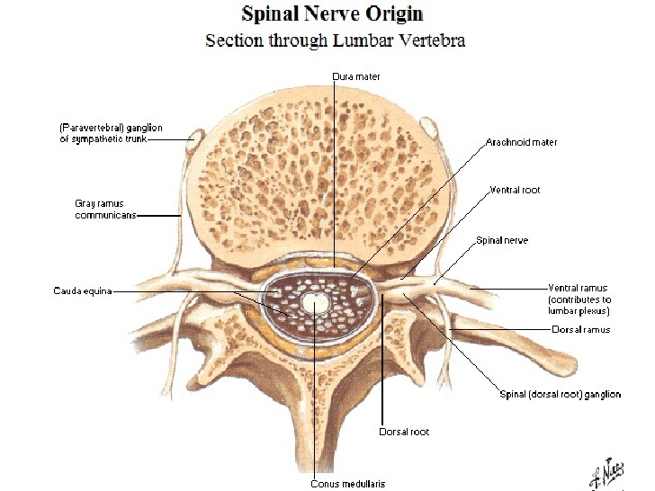
~the spinal cord gives attachment ,on either side,to a series of spinal nerves.
~each spinal nerve arises by two roots:-
1.anterior (or ventral)
2.posterior(or dorsal)
~each root is made up of a number of rootlets.the length of the spinal cord giving origin to the rootlets for one spinal nerve constitutes one spinal segment.
~as the spinal cord is much shorter than the length of the vertebral column the spinal segments do not lie opposite the corresponding vertebrae.
~in estimating the position of a spinal segment in relation to the surface of the body it is important to remember that a vertebral spine is always lower than the corresponding spinal segment.
~as a rough guide it may be stated that in the cervical region there is a difference of one segment,in the upper thoracic region there is a difference of two segment ,and in the lower thoracic region there is a difference of three segments.
Tracts of the spinal cord:-
~A collection of nerve fibers that connects two masses of grey matter within the central nervous system is called a tract.tracts may be ascending or descending.
~they are usually named after the masses of grey matter connected by them.some tracts are called fasciculi or lemnisci.
*Lateral spinothalamic tract:-
~Pain & T
*Anterior spinothalamic tract:-
~Light(crude) touch & pressure
*Fasciculus cuneatus
*Fasciculus gracilis
~discriminatory touch,vibration,info from muscles & joints.
*Anterior spinocerebellar tract
*Posterior spinocerebellar tract
~unconscious info from muscles,joints,skin,subcut
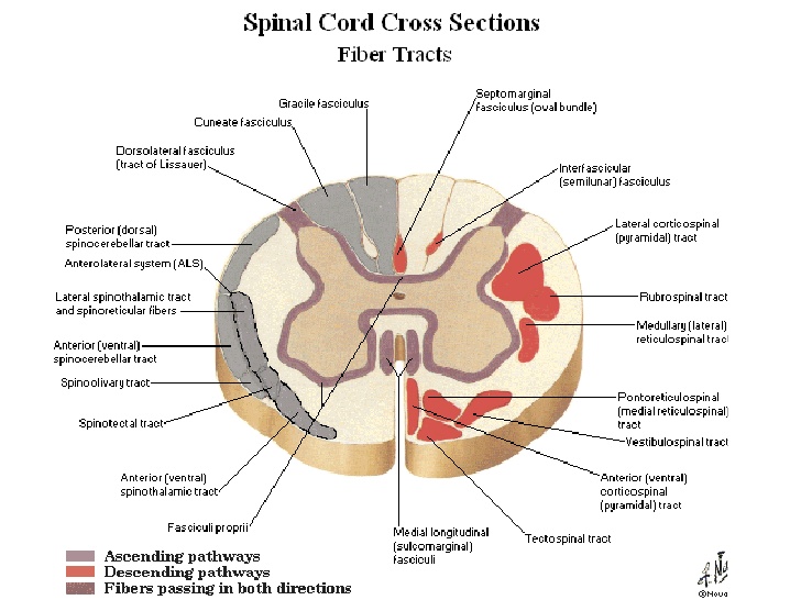
*Spinotectal tract:-
~spinovisual reflexes
*spinoreticular tract:-
~info from muscles, joints & skin to the reticular formation
*spino-olivary tract:-
~indirect pathway to cerebellum
*Lateral spinothalamic tract:-
~pain & temp pathways
*1st order neurons:-
*pain conducted by A type fibers & C type fibers
*2nd order neurons:-
~deccussate to the opposite side
~ends in thalamus(ventral posterolateral nucleus)
*3rd order neurons:-
~ends in sensory area in postcentral gyrus
*Anterior spinothalamic tract:-
~light (crude) touch & pressure pathways
*Posterior white column:-
~discriminative touch,vibratory sense,consious muscle joint sense(conscious proprioception)
*Posterior spinocerebellar tract:-
~muscle joint sense pathways to cerebellum.
~unconscious proprioception
~muscle joint info from muscle spindles,GTO,joint receptors of the trunk & lower limbs.
~info is used by the cerebellum in the coordination of movements & maintenance of posture.
*Anterior spinocerebellar tract:-
~majority of 2nd order neurons cross to the opposite side.
~enter cerebellum through superior cerebellar peduncle.
~info from trunk ,upper & lower limbs.
~also carries info from skin & subcut tissue.
*Descending tracts:-
~upper motor neurons
~lower motor neurons
~corticospinal tracts:-
~concerned with voluntary,discrete,skilled movements.
*Reticulospinal tract:-
~facilitates or inhibits voluntary movement or reflex activity.
*Tectospinal tract:-
~reflex postural movements in response to visual stimuli.
*Rubrospinal tract:-
~fascilitates activity of flexor muscles & inhibits activity of extensor muscles.
*Vestibulospinal tract:-
~fascilitates extensor muscles,inhibits flexor muscles.
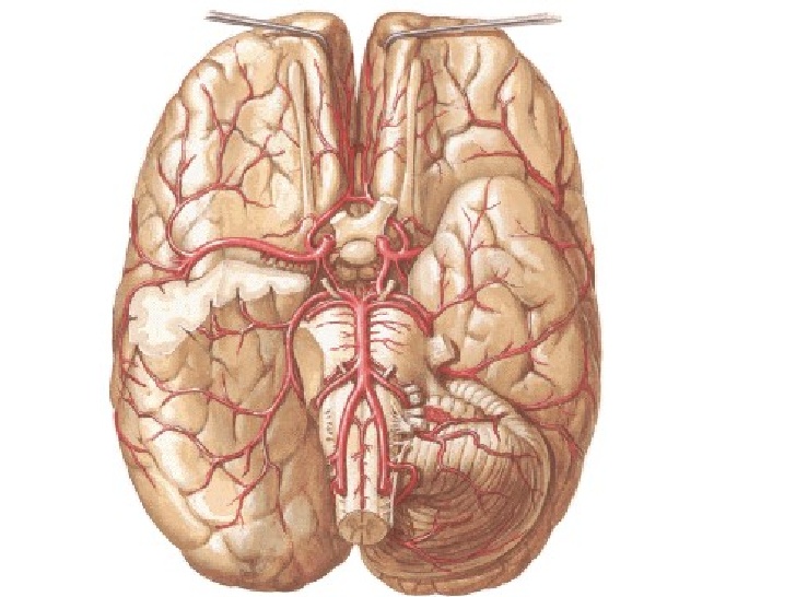
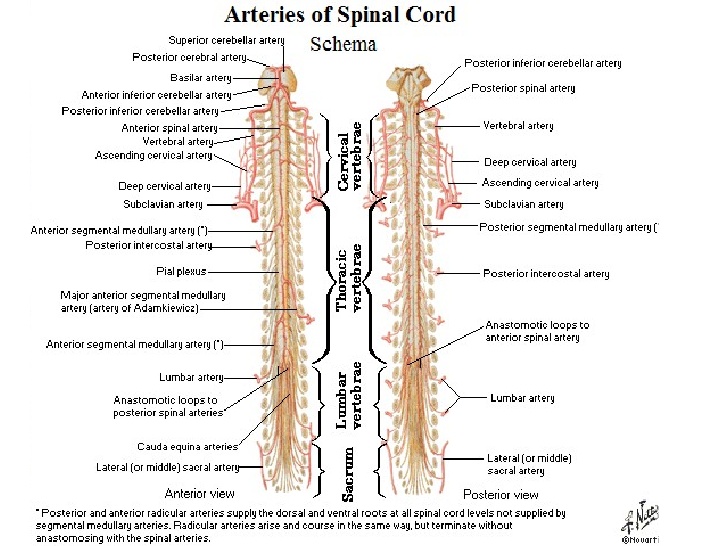
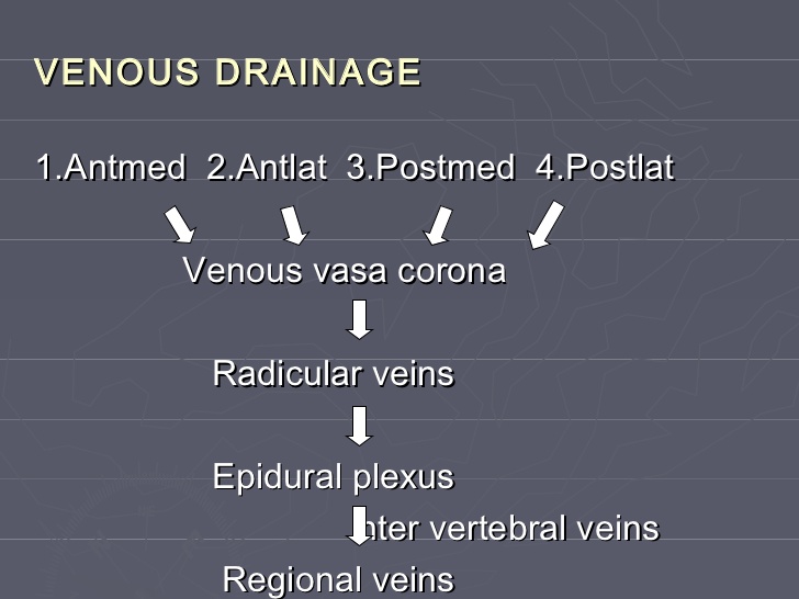
Blood supply:-
*Arteries of spinal cord:-
1.anterior spinal artery
2.posterior spinal artery
3.segmental spinal arteries
*Anterior spinal artery:-
~formed by the union of 2 arteries.
~from vertebral artery
~supply anterior 2/3 of spinal cord.
*Posterior spinal arteries:-
~arises from vertebral artery or posterior inferior cerebellar arteries(PICA)
~Descend close to the posterior roots.
~supply posterior 1/3 of spinal cord.
*Segmental spinal arteries:-
~Branches of arteries outside the vertebral column.
~gives off the anterior & posterior radicular arteries.
~great anterior medullary artery of adamkiewicz.
~arise from lateral intercostal artery or lumbar artery at any level from T8-L3
*main source of blood is the vertebral arteries…cervical
~radicular arteries ..spinal branch of vertebral ,asc cervical,deep cervical ,inercostal,lumbar and sacral art
~arteria radicularis magna
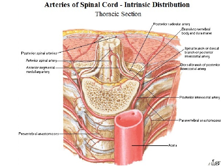

*Distribution of blood supply:-
~radicular art:-grey & white matter
~anterior spinal :-ant two-third ventral grey matter,part of dorsal grey matter,ant and lat feniculi
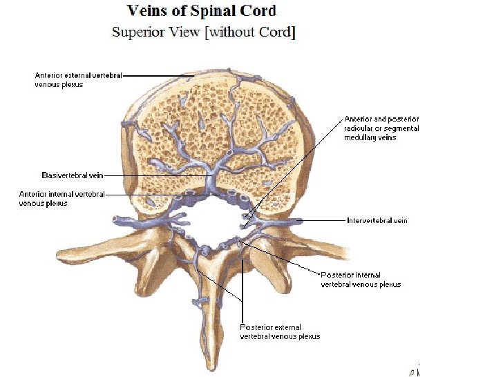
~posterior spinal:-post one third posterior horn and posterior feniculus
Meninges:
*Dura mater:-
~dense,strong fibrous membrane.
~encloses the spinla cord & cauda equina
~continous above with the meningeal layer of dura covering the brain.
~ends at the level of S2.
~separated from wall of vertebral canal by the extradural space
~contains loose areolar tissue & internal vertebral venous space
*Arachnoid mater:-
~delicate impermeable membrane.
~lies between pia and dura mater.
~separated from pia mater by subarchanoid space.
~continous above with archanoid mater covering the brain.
~ends on filum terminale at level of S2.
*Pia mater:-
~vascular membrane
~closely covers spinal cord
~thickened on either side between nerve roots to form the ligamentum denticulatum
Clinical importance:-
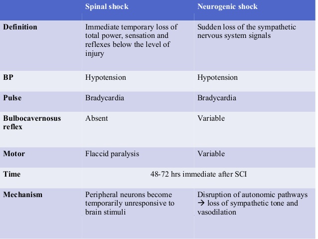
*Spinal shock:-
~follows acute severe damage to the spinal cord.
~all cord functions below the level of the lesion become depressed lost.
~sensory impairment and flaccid paralysis occur.
~segmental spinal reflexes are depressed
~persists for less than 24 hours (may be as long as 1-4 weeks)
*Poliomyelitis:-
~acute viral infection of the neurones of anterior gray column.
~motor nuclei of cranial nerves.
~death of motor neurons cells-paralysis & wasting of muscles.
~muscles of lower limb more often affected.
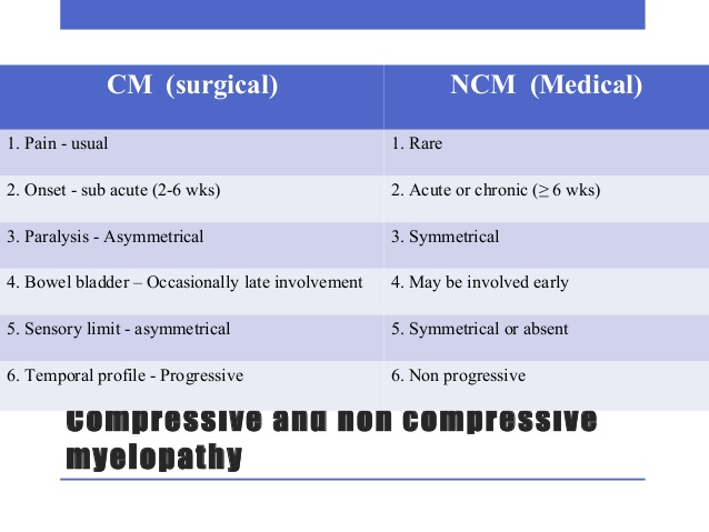
*Spinal cord pathology:-
*Vertebral cause:-
1.trauma
2.disc prolapse
3.tumor-primary,eg-MM,secondary-eg.breast,thyroid,prostate,bronchus
4.spinal TB-(potts diease)
*Meningeal cause:-
1.epidural abcess
2.tumor-meningioma,neurofibroma,lymphoma,leukaemia
*Spinal cord itself:-
*Developmental:-
~syringomyelia
~meningomyelocoele
~tetherd cord syndrome
*Degenerative:-
~MND
~FA
~SCD
~HSP
*Demyelinating/inflammatory:-
~transverse myelitis
~multiple sclerosis
~neuromyelitis optica
*Infective:-
~bacterial-TB,Syphilis
~viral-EBV,polio,HIV,VZV,HSV
~parasitic-schistosomiasis,toxoplasmosis
*Deficiency:-
~vitamin B12 Deficiency
~vitamin E deficiency
~copper deficiency
~lathyrism
*Vascular:-
~vasculitis
~infraction
~haemorhage
~AVM
*Physcial agents:-
~radiation
~lightening injury
*Paraneoplastic
Localization of spinal cord diease:–
~presence of horizontally defined level below which there will be impairment of sensory,motor,and autonomic function.
*Cervical cord:-
~above C5-spastic qaudriplegia,and diaphgram weakness
~C5-T1:-(qaudriplegia,(LMN signs and segmental sensory loss in the arms & UMN signs in the legs)& respiratory(intercostal) muscle weakness.
~at C5-C6:-loss of power & reflex of biceps
~at C7:-weakness in fingers & wrist extensors & triceps.
~at C8:-finger & Wrist flexion are impaired.
~horners syndrome may accompany
*Thoracic cord:-
~spastic paraplegia with a sensory level on the trunk.
~bowel & bladder involvement
~abdominal reflex(T8-T12)lost above T8 lesion
(segmental lesion T8-T9:above the umbilicus)
(T10-T12:Below the umbilicus)
*Lumbar cord:-
~L2-L4:-
weakness of flexion & adduction of thigh
weakness in leg extension at knee
absent knee jerks(L3-L4)
*L5-S1:-
~weakness of foot & ankle and flexion at the knee & extension of the thigh
~absent ankle jerks(S1)
*Sacral cord:-
~saddle anesthesia(S3-S5)
~prominent bowel & bladder dysfunction & impotence.
~absent bulbocavernous (S2-S4) & anal reflex(S4-S5)
Types of spinal cord lesion:-
*complete or transverse lesion
*Incomplete lesion:-
~anterior cord syndrome
~posterior cord syndrome
~hemi cord syndrome
~central cord syndrome
~foramen magnum syndrome
~conus medullaris syndrome
~cauda equina syndrome
*Complete cord transection syndrome:-
~bilateral spastic(paraparesis/quadriparasis)
~bilateral loss of all moodalities of sensation
~bowel & bladder dysfunction
~LMN feature at the level of lesion
~cause:-trauma,vasculitis
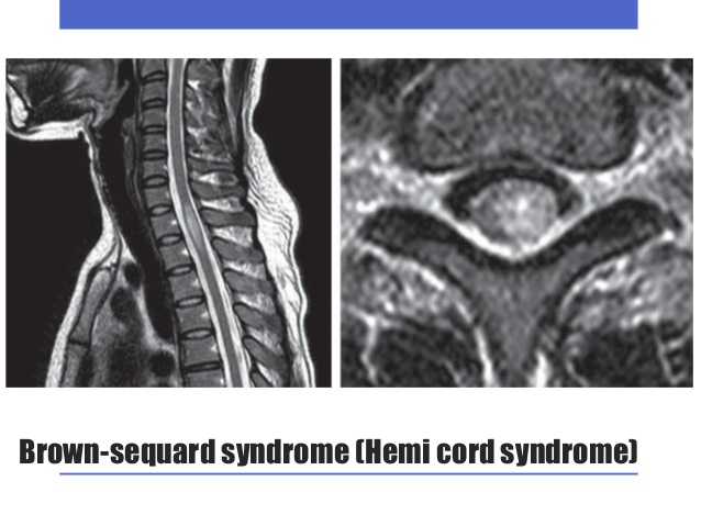
*Brown sequard syndrome:-
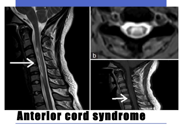
*Anterior cord syndrome:-
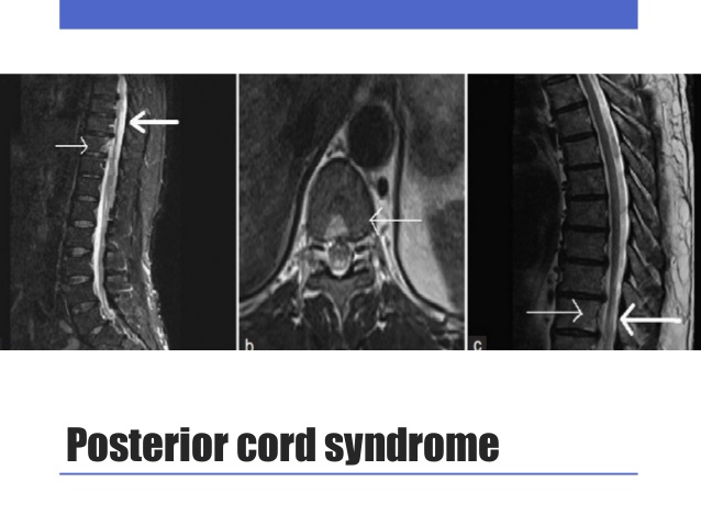
*Posterior cord syndrome:-
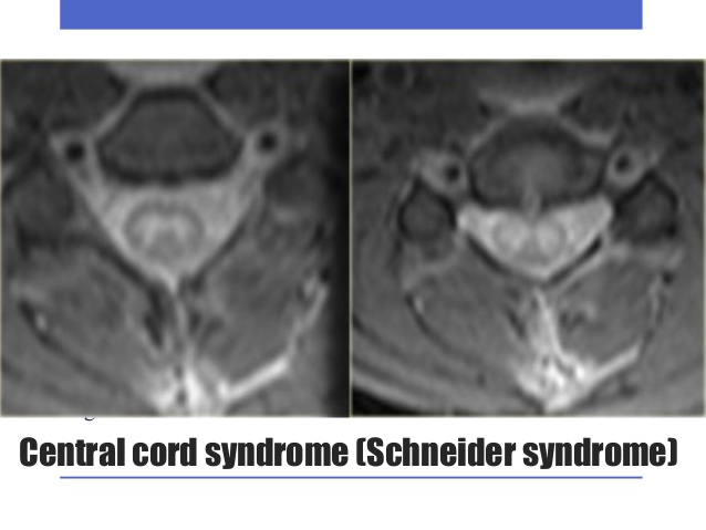
*Central cord syndrome:-
*Foramen magnum syndrome:-
*C/F:-
~neck pain-radiating to shoulder
~occipital H/A
~variable sensory loss
~weakness & wasting of hand & neck muscles
~qaudriparesis(round the clock)
*Cause:-
~compressive lesion(meningioma,neurofibroma)in the region of foramen magnum
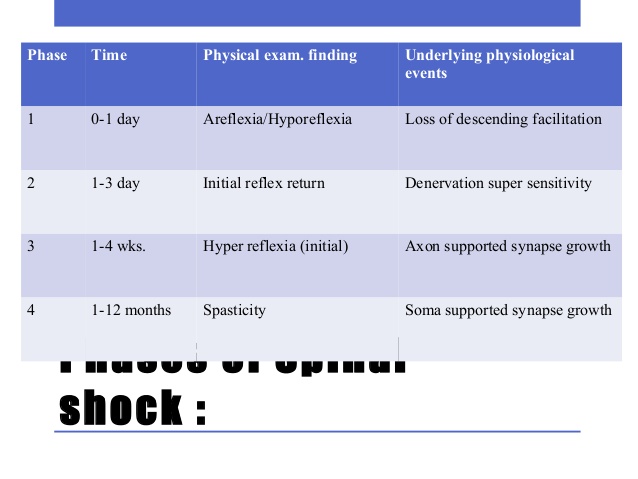
*Spinal shock syndrome:-
~this clinical condition follows acute severe damage to the cord.
~all cord function below the level of lesion becomes depressed or lost.
~usually last less than 24 hrs bt may last for 4-6 weeks.
~on recovery:-reflex-power-tone may regain this fashion
~5-10% patients may not recover from spinal shock.
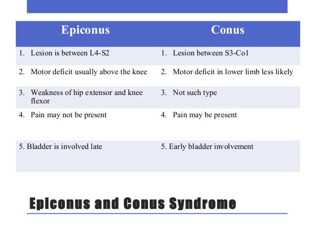
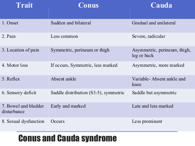
*Conus medullaris syndrome:-
~bilateral saddle anesthesia
~prominent bowel & bladder dysfunction(urinary retention & anal incontinence)
~impotence
~absent anal reflex
~radicular low back pain
~asymmetrical lower limb weakness & sensory loss
~variable areflexia
~relative sparing of bowel & bladder
~planter may be flexor or absent
*cause:-
~disc prolapse
~tumor
~trauma
20 Comments