
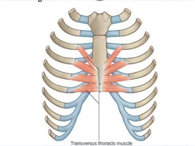
Transversus thoracis muscle
Transversus thoracis muscle Anaomy
Transversus thoracis muscle lies internal to the thoracic cage, anteriorly. It is a thin plane of muscular and tendinous fibers, situated upon the inner surface of the front wall of the chest.
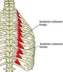
On the inside surface of the anterior chest wall, there is a muscle called transversus thoracis (also known as triangularis sternae or sternocostalis). Along with the intercostals, subcostals, levatores costarum, and serratus posterior muscles, it is one of the intrinsic muscles of the chest wall.
Transversus thoracis, like all the intrinsic chest muscles (with the exception of levatores costarum), is innervated by the nearby intercostal nerves. Transversus thoracis aids in supporting the thoracic cage and moving the ribs during forced breathing, as do all of these muscles.
Origin:
Costal cartilages of last 3-4 true ribs, body of sternum and xiphoid process.
Insertion:
Ribs/costal cartilages 2-6 .
Nerve Supply:
Intercostal nerves.
Actions:
Depresses ribs.
An secondary respiratory muscle called the transversus thoracis is engaged during forced expiration. During forced expiration, it pulls ribs 2 through 6 towards the sternum, depressing those ribs. As a result, the thoracic cavity’s anteroposterior diameter is reduced.
Additionally, the transversus thoracis stiffens the thoracic wall during inspiration, preventing the chest wall from moving paradoxically.
Relations
The anterior wall of the anterior mediastinum is formed by the transversus thoracis, which is positioned deep to the centre of the thoracic cage. It divides the intercostal nerves from the pleura. The transversus thoracis’ inferior border and the transversus abdominis’ upper border are in close proximity to one another. Prior to both muscles, the superior epigastric artery and vein pass.
Clinical Importance
Due to its proximity to the internal thoracic artery, also known as the internal mammary artery (IMA) in clinical terminology, the transversus thoracis plays a significant role in heart surgery. This artery is especially well-suited as a transplant intermediary for a coronary artery bypass because of how it exits the subclavian artery and how close it is to the heart. Additionally, just one side needs to be anastomosed.
Since this is typically the easiest location to locate and dissect the vessel, surgeons prefer to start the artery harvesting procedure between the first rib and the highest insertion tendon of the transversus thoracis. The transversus thoracis muscle fibres surround the internal thoracic artery more caudally.
3 Comments