Subtalar Osteoarthritis
Table of Contents
What is Subtalar Osteoarthritis?
Subtalar osteoarthritis is a degenerative joint condition that affects the subtalar joint, which is located between the talus (ankle bone) and calcaneus (heel bone).
Osteoarthritis is a common form of arthritis characterized by the gradual breakdown of the cartilage within a joint. The subtalar joint plays a crucial role in foot and ankle movement, contributing to inversion and eversion.
Repetitive trauma or ankle sprains often lead to subtalar osteoarthritis, which impairs the patient’s quality of life. Up to 78% of those with chronic ankle instability may develop posttraumatic ankle osteoarthritis.
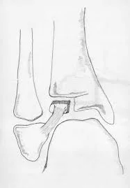
The subtalar joint is the junction that connects the calcaneus and the talus. It is a complex joint that is essential to propulsion and stress absorption in human movement. This joint is designed to provide the foot with either a rigid propulsive framework or a flexible shock-absorbing structure.
When the subtalar joint is everted, or in valgus, the transverse tarsal joints become freed, resulting in a flexible foot structure. When the subtalar joint inverts, the transverse tarsal joints lock, producing a rigid lever arm that aids in movement.
The sinus tarsi divides the subtalar joint into the talocalcaneonavicular joint and the talocalcaneal joint. This finding explains why only the posterior subtalar facet is visible from the more conventional surgical techniques that do not penetrate the sinus tarsi.
Symptoms of subtalar osteoarthritis:
- Pain
- Swelling
- Stiffness
- Joint deformity
Causes of Subtalar Osteoarthritis:
Subtalar arthritis is a disorder affecting the joint between the foot and ankle. It happens when the joint sustains severe wear and tear from an accident or excessive usage. There is often no identified cause for persistent lateral ankle pain. On the other hand, an injury or even a particular pair of shoes can be the cause of the incident.
Due to its extensive range of motion, the subtalar joint is prone to injury. When you walk, your feet are constantly rising and falling. This puts a lot of tension on the joints in your feet, which over time might lead to arthritis. If you have subtalar arthritis, getting up from a sitting position may cause you pain.
Arch support can help someone with subtalar joint arthritis and an uneven foot arch to properly align their forefoot and heel. Restricting aberrant motion and using insoles, ankle, and foot supports can help reduce discomfort. If the ankle does not heal properly, the bones may rub against one another and cause pain.
Diagnosis
Diagnosis typically involves a thorough clinical examination, medical history review, and imaging studies such as X-rays.
Physical examination:
Clinical signs of pain and dysfunction may appear years or even decades after the original harm. Patients usually complain of instability, stiffness, or edema that initially improves with rest, as well as pain in the ankles and hindfoot that worsens with weight-bearing or activity. Typically, symptoms begin gradually and worsen with time.
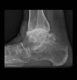
Imaging of subtalar osteoarthritis:
Examples of imaging evaluations that need to be tailored to specific clinical settings include magnetic resonance imaging, ultrasound assessment, computed tomography (which can be performed using a standard or weight-bearing approach), weight-bearing radiography (which uses both standard and specialty views), and magnetic resonance imaging.
Ankle radiography is typically the first imaging technique to be performed. The majority of medical practitioners radiograph the entire leg, including the foot, while standing. Some could ask for axial or Salzman viewpoints.
A computed tomographic scan is useful since it evaluates the degree of the malformation in addition to ruling out a coalition. Due to its ability to identify the internal fixation of pain, single-photon emission CT has become more and more popular in recent years.
This is especially true in posttraumatic scenarios where standard imaging modalities may not be able to determine the pain’s origin. Since plain films do not fully characterize non-ossified components such as articular cartilage, marrow tissue, and synovial fluid, it is possible to miss any early symptoms of degradation.
Magnetic resonance imaging is an additional option if needed. It might be difficult to detect joint space narrowing or degeneration on routine standing radiographs because of the complex shapes of the bones and joints without the use of specialized
Treatment of Subtalar Osteoarthritis:
Treatment approaches for subtalar osteoarthritis aim to alleviate symptoms, improve joint function, and enhance overall mobility. Conservative measures may include medications, physical therapy, and orthotic devices. In more advanced cases, surgical interventions such as arthrodesis (joint fusion) or joint replacement may be considered.
Reducing the rear foot’s range of motion should be the aim of nonoperative treatment to minimize pain. Weight loss, medications, subtalar joint injections, shock-absorbing materials like shoes or insoles, and specialized insoles made to limit hindfoot motion are examples of traditional conservative therapy.
By reducing the hindfoot’s propensity to evert, insoles with a central post can help ease subtalar pain. Shoes with a rocker bottom have become more and more fashionable in the last 10 years.
An intraarticular injection of glucocorticoids, with or without anesthetic, is a useful treatment for osteoarthritis in the ankle and various kinds of arthritis throughout the body. This therapy strategy is commonly utilized when more conservative approaches are ineffective for treating subtalar osteoarthritis.
The wide range of reported efficacy in post-traumatic ankle arthritis is usually accounted for by the 30 to 80% success rate of needle insertion when manual palpation is the only means of guidance. By using ultrasonography as guidance during injections, the number of errors resulting from human error has decreased, and the success rate of placing needles has increased from 32 to 97%.
Injections into the subtalar joint may be challenging because of the anatomy of the foot and ankle. Among the three that have been described and are often used are the posteromedial, posterolateral, and anterolateral approaches. The posterolateral approach is the most commonly used method. When using the posterolateral approach, the injection can be given using a long-axis technique that provides continuous visualization of the whole needle shaft and tip.
Post-Operative Course:
Day 1
- The foot is elevated, cooled, and taking pain medication while being wrapped in a heavy bandage and splint.
- You should anticipate soreness and foot numbness for 12 to 24 hours.
- Be prepared for bloody drainage.
- Don’t take off the bandage.
Day 14
- Radiographs are taken, a fresh dressing is applied, and a boot is put on.
- Don’t lift anything for about a week or two.
6 weeks
- After the boot is taken off, X-rays are taken.
- Proceed as usual within the boot, then progressively increase your weight bearing by utilizing your level of pain as a guide.
12 weeks
- Start a fitness routine.
- Orders for orthotic arch supports are often placed.
Physiotherapy treatment of Subtalar Osteoarthritis:
- Pain and swelling can be reduced after exercise by elevating the leg and placing an ice pack on the ankle and rear of the foot.
- By adding a small heel lift or cushion to your shoe, you might be able to take lengthy walks without limping and relieve some of the tension on your tense ankle.
- Since it stops the subtalar joint from moving too much, an ankle brace—such as a strap around the ankle—or even tying the ankle to the backfoot may be beneficial.
- Get a new pair of comfortable shoes and stuff them full of cushioning. As a result, the ankles might not be stressed.
- Physical therapy or physiotherapy are effective ways to relieve discomfort associated with subtalar arthritis. It also provides stability to the foot, helps restore lost range of motion, and tones the muscles surrounding the ankle.
- Using an assistance device, such as a cane or walker, could be helpful if the discomfort worsens. It would be advantageous to decrease the load on the joint to ease the strain and pain in the affected area.
General Factors of Postoperative Recovery:
- You won’t be able to walk on the leg for at least six weeks. Bone healing and more X-rays will provide an exact time frame.
- To keep off your feet, you’ll need to utilize a wheelchair, walker, crutches, or rollout equipment.
- The limb will spend two weeks in a rigid plaster cast following surgery.
- You’ll have your first follow-up visit to have the sutures taken out in around two weeks.
- We frequently attach a removable boot for you to wear for about two weeks.
- After your left ankle surgery, after two weeks, you should be ready to drive an automated vehicle. If everything goes according to plan, you can get back behind the wheel in three to four weeks.
- You may begin placing some weight on the boot at around six weeks, depending on how much discomfort you are experiencing and the instructions you have been given.
- Physical therapy helps restore the ankle’s strength and range of motion.
- Schedule a one- to two-month appointment with a physical therapist.
- There will be some swelling in the foot, ankle, and leg for around six months.
- You will continue to gain strength and mobility for over a year after the treatment.
- You should expect to be sore and uncomfortable for four to six months following surgery.
- Orthotic arch supports may be helpful during the recovery process.
Summary:
Repetitive trauma or ankle sprains often lead to subtalar osteoarthritis, which impairs the patient’s quality of life. Up to 78% of those with chronic ankle instability may develop posttraumatic ankle osteoarthritis.
The subtalar joint is the junction that connects the calcaneus and the talus. It is an intricate joint that is essential to propulsion and stress absorption in human movement. This joint is designed to provide the foot with either a rigid propulsive framework or a flexible shock-absorbing structure.
When the subtalar joint is everted, or in valgus, the transverse tarsal joints become freed, resulting in a flexible foot structure. When the subtalar joint inverts, the transverse tarsal joints lock, producing a rigid lever arm that aids in movement. The sinus tarsi divides the subtalar joint into the talocalcaneonavicular joint and the talocalcaneal joint. This finding explains why only the posterior subtalar facet is visible from the more conventional surgical techniques that do not penetrate the sinus tarsi.
FAQ
The joint in the backfoot under the ankle joint is called the subtalar joint, and it can be impacted by a kind of arthritis called subtalar arthritis. The condition’s defining feature is uncomfortable hindfoot discomfort, which is exacerbated by standing and walking, especially on uneven surfaces.
Subtalar arthritis is the name for arthritis affecting the cartilage of the subtalar joint. The most common cause of subtalar arthritis is a traumatic injury to the hindfoot, which is often seen after a talus or calcaneus fracture. Inflammation and pain in the rearfoot are signs of subtalar arthritis.
While there isn’t a known cure for arthritis, there are several treatment options that can slow the disease’s course and minimize symptoms. With the right medicine, many people with arthritis may live happy, active lives, and manage their pain.
Subtalar joint diseases can cause problems with gait and mobility, particularly when a person is walking on uneven ground. If left untreated, damage to the subtalar joint may become irreparable.
Osteoarthritis is an incurable chronic condition, but it doesn’t necessarily become worse with time—in fact, in certain situations, it may even get better. To reduce the symptoms, there are also several treatments available. On rare occasions, simple remedies like consistent exercise might be utilized to cure minor problems.